Daniel Adrian Silva Souza, Fábio Wildson Gurgel Costa, Diego Santiago de Mendonça, Esther Carneiro Ribeiro, Paulo Goberlânio de Barros Silva, Frederico Sampaio Neves
{"title":"上颌窦发育不良及相关解剖变异的计算机断层扫描评估:全球证据的系统回顾和荟萃分析。","authors":"Daniel Adrian Silva Souza, Fábio Wildson Gurgel Costa, Diego Santiago de Mendonça, Esther Carneiro Ribeiro, Paulo Goberlânio de Barros Silva, Frederico Sampaio Neves","doi":"10.1007/s11282-023-00726-2","DOIUrl":null,"url":null,"abstract":"<p><strong>Objective: </strong>To summarize the scientific evidence on the prevalence of maxillary sinus hypoplasia (MSH) and associated anatomical variations as assessed by computed tomography scans.</p><p><strong>Study design: </strong>This PROSPERO-registered systematic review followed the recommendations of the PRISMA guidelines. Search algorithms were constructed for each of the six databases and gray literature. After screening the references (Rayyan<sup>®</sup>), the extracted data were meta-analyzed according to a random-effects model. The joanna briggs critical appraisal tool assessed the methodological quality of the included studies. The GRADE approach was used to estimate the certainty of the evidence.</p><p><strong>Results: </strong>From a total of 2781 studies screened, 22 were considered for four meta-analysis. The prevalence of MSH in 7358 patients was 5.65% (CI95% = 4.07-7.47%) with significant heterogeneity between studies (p < 0.001, I<sup>2</sup> = 89.30%). MSH was identified in 295 patients, of whom 82.38% (CI95% = 75.82-88.09%) had unilateral hypoplasia and 17.62% (CI95% = 11.91-24.18%) bilateral hypoplasia with moderate heterogeneity between studies (p < 0.0503, I<sup>2</sup> = 42.87%). The prevalence of MSH in 9998 maxillary sinuses was 3.77% (95% CI = 2.44-5.38%), with significant heterogeneity between studies (p < 0.001, I<sup>2</sup> = 92.84%). Hypoplastic/aplastic uncinate process, concha bullosa and paradoxical concha were the most reported anatomical variations. The studies presented a low-moderate methodological quality. The certainty of the evidence was very low to moderate.</p><p><strong>Conclusion: </strong>The prevalence of maxillary sinus hypoplasia observed was 5.65%, with most cases being unilateral.</p>","PeriodicalId":56103,"journal":{"name":"Oral Radiology","volume":" ","pages":"124-137"},"PeriodicalIF":1.6000,"publicationDate":"2024-04-01","publicationTypes":"Journal Article","fieldsOfStudy":null,"isOpenAccess":false,"openAccessPdf":"","citationCount":"0","resultStr":"{\"title\":\"Computed tomography assessment of maxillary sinus hypoplasia and associated anatomical variations: a systematic review and meta-analysis of global evidence.\",\"authors\":\"Daniel Adrian Silva Souza, Fábio Wildson Gurgel Costa, Diego Santiago de Mendonça, Esther Carneiro Ribeiro, Paulo Goberlânio de Barros Silva, Frederico Sampaio Neves\",\"doi\":\"10.1007/s11282-023-00726-2\",\"DOIUrl\":null,\"url\":null,\"abstract\":\"<p><strong>Objective: </strong>To summarize the scientific evidence on the prevalence of maxillary sinus hypoplasia (MSH) and associated anatomical variations as assessed by computed tomography scans.</p><p><strong>Study design: </strong>This PROSPERO-registered systematic review followed the recommendations of the PRISMA guidelines. Search algorithms were constructed for each of the six databases and gray literature. After screening the references (Rayyan<sup>®</sup>), the extracted data were meta-analyzed according to a random-effects model. The joanna briggs critical appraisal tool assessed the methodological quality of the included studies. The GRADE approach was used to estimate the certainty of the evidence.</p><p><strong>Results: </strong>From a total of 2781 studies screened, 22 were considered for four meta-analysis. The prevalence of MSH in 7358 patients was 5.65% (CI95% = 4.07-7.47%) with significant heterogeneity between studies (p < 0.001, I<sup>2</sup> = 89.30%). MSH was identified in 295 patients, of whom 82.38% (CI95% = 75.82-88.09%) had unilateral hypoplasia and 17.62% (CI95% = 11.91-24.18%) bilateral hypoplasia with moderate heterogeneity between studies (p < 0.0503, I<sup>2</sup> = 42.87%). The prevalence of MSH in 9998 maxillary sinuses was 3.77% (95% CI = 2.44-5.38%), with significant heterogeneity between studies (p < 0.001, I<sup>2</sup> = 92.84%). Hypoplastic/aplastic uncinate process, concha bullosa and paradoxical concha were the most reported anatomical variations. The studies presented a low-moderate methodological quality. The certainty of the evidence was very low to moderate.</p><p><strong>Conclusion: </strong>The prevalence of maxillary sinus hypoplasia observed was 5.65%, with most cases being unilateral.</p>\",\"PeriodicalId\":56103,\"journal\":{\"name\":\"Oral Radiology\",\"volume\":\" \",\"pages\":\"124-137\"},\"PeriodicalIF\":1.6000,\"publicationDate\":\"2024-04-01\",\"publicationTypes\":\"Journal Article\",\"fieldsOfStudy\":null,\"isOpenAccess\":false,\"openAccessPdf\":\"\",\"citationCount\":\"0\",\"resultStr\":null,\"platform\":\"Semanticscholar\",\"paperid\":null,\"PeriodicalName\":\"Oral Radiology\",\"FirstCategoryId\":\"3\",\"ListUrlMain\":\"https://doi.org/10.1007/s11282-023-00726-2\",\"RegionNum\":3,\"RegionCategory\":\"医学\",\"ArticlePicture\":[],\"TitleCN\":null,\"AbstractTextCN\":null,\"PMCID\":null,\"EPubDate\":\"2023/12/11 0:00:00\",\"PubModel\":\"Epub\",\"JCR\":\"Q3\",\"JCRName\":\"DENTISTRY, ORAL SURGERY & MEDICINE\",\"Score\":null,\"Total\":0}","platform":"Semanticscholar","paperid":null,"PeriodicalName":"Oral Radiology","FirstCategoryId":"3","ListUrlMain":"https://doi.org/10.1007/s11282-023-00726-2","RegionNum":3,"RegionCategory":"医学","ArticlePicture":[],"TitleCN":null,"AbstractTextCN":null,"PMCID":null,"EPubDate":"2023/12/11 0:00:00","PubModel":"Epub","JCR":"Q3","JCRName":"DENTISTRY, ORAL SURGERY & MEDICINE","Score":null,"Total":0}
Computed tomography assessment of maxillary sinus hypoplasia and associated anatomical variations: a systematic review and meta-analysis of global evidence.
Objective: To summarize the scientific evidence on the prevalence of maxillary sinus hypoplasia (MSH) and associated anatomical variations as assessed by computed tomography scans.
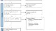
Study design: This PROSPERO-registered systematic review followed the recommendations of the PRISMA guidelines. Search algorithms were constructed for each of the six databases and gray literature. After screening the references (Rayyan®), the extracted data were meta-analyzed according to a random-effects model. The joanna briggs critical appraisal tool assessed the methodological quality of the included studies. The GRADE approach was used to estimate the certainty of the evidence.
Results: From a total of 2781 studies screened, 22 were considered for four meta-analysis. The prevalence of MSH in 7358 patients was 5.65% (CI95% = 4.07-7.47%) with significant heterogeneity between studies (p < 0.001, I2 = 89.30%). MSH was identified in 295 patients, of whom 82.38% (CI95% = 75.82-88.09%) had unilateral hypoplasia and 17.62% (CI95% = 11.91-24.18%) bilateral hypoplasia with moderate heterogeneity between studies (p < 0.0503, I2 = 42.87%). The prevalence of MSH in 9998 maxillary sinuses was 3.77% (95% CI = 2.44-5.38%), with significant heterogeneity between studies (p < 0.001, I2 = 92.84%). Hypoplastic/aplastic uncinate process, concha bullosa and paradoxical concha were the most reported anatomical variations. The studies presented a low-moderate methodological quality. The certainty of the evidence was very low to moderate.
Conclusion: The prevalence of maxillary sinus hypoplasia observed was 5.65%, with most cases being unilateral.
期刊介绍:
As the official English-language journal of the Japanese Society for Oral and Maxillofacial Radiology and the Asian Academy of Oral and Maxillofacial Radiology, Oral Radiology is intended to be a forum for international collaboration in head and neck diagnostic imaging and all related fields. Oral Radiology features cutting-edge research papers, review articles, case reports, and technical notes from both the clinical and experimental fields. As membership in the Society is not a prerequisite, contributions are welcome from researchers and clinicians worldwide.

 求助内容:
求助内容: 应助结果提醒方式:


