{"title":"猴子和人类后脾神经细胞学的比较","authors":"Brent A. Vogt, Douglas L. Rosene","doi":"10.1002/cne.25561","DOIUrl":null,"url":null,"abstract":"<p>Retrosplenial cortex (RSC) has unique problems for human neuroimaging studies as its divisions are small, at the lower end of functional scanner spatial resolution, and it is buried in the callosal sulcus. The present study sought to define the cytoarchitecture of RSC in human and monkey brains along its entire anteroposterior extent. The results show anterior extensions, a newly defined dichotomy of area 30, a new area p30, and an area p29v in monkey that differentiates into three divisions in human. Accordingly, anterior (a), intermediate (i), and posterior (p) divisions of areas 29l, 29m, 30l, and 30m were identified. Posterior area 29 has higher neuron packing in the granular layer than anterior and intermediate divisions of area 29. A newly detected dysgranular area p30 has larger neurons in layers II–IIIab than a30 and i30 and with substantially higher NFP expression in layer IIIab of posterior areas than areas a30 and i30. Medial area 30 has larger pyramids and higher NFP expression in all layers than area 30l. The new area p30 was seen between areas p29m and p30I in both species. Finally, a ventral area p29v is present in monkeys. This latter area appears to differentiate into three divisions in human with the most extensive granular layer adjacent to layer I in p29vm and p29vl. Functional imaging has identified pRSC as part of a cognitive map which is engaged in spatial navigation and localization of personally relevant objects.</p>","PeriodicalId":15552,"journal":{"name":"Journal of Comparative Neurology","volume":"531 18","pages":"2044-2061"},"PeriodicalIF":2.3000,"publicationDate":"2023-12-07","publicationTypes":"Journal Article","fieldsOfStudy":null,"isOpenAccess":false,"openAccessPdf":"","citationCount":"0","resultStr":"{\"title\":\"Comparison of monkey and human retrosplenial neurocytology\",\"authors\":\"Brent A. Vogt, Douglas L. Rosene\",\"doi\":\"10.1002/cne.25561\",\"DOIUrl\":null,\"url\":null,\"abstract\":\"<p>Retrosplenial cortex (RSC) has unique problems for human neuroimaging studies as its divisions are small, at the lower end of functional scanner spatial resolution, and it is buried in the callosal sulcus. The present study sought to define the cytoarchitecture of RSC in human and monkey brains along its entire anteroposterior extent. The results show anterior extensions, a newly defined dichotomy of area 30, a new area p30, and an area p29v in monkey that differentiates into three divisions in human. Accordingly, anterior (a), intermediate (i), and posterior (p) divisions of areas 29l, 29m, 30l, and 30m were identified. Posterior area 29 has higher neuron packing in the granular layer than anterior and intermediate divisions of area 29. A newly detected dysgranular area p30 has larger neurons in layers II–IIIab than a30 and i30 and with substantially higher NFP expression in layer IIIab of posterior areas than areas a30 and i30. Medial area 30 has larger pyramids and higher NFP expression in all layers than area 30l. The new area p30 was seen between areas p29m and p30I in both species. Finally, a ventral area p29v is present in monkeys. This latter area appears to differentiate into three divisions in human with the most extensive granular layer adjacent to layer I in p29vm and p29vl. Functional imaging has identified pRSC as part of a cognitive map which is engaged in spatial navigation and localization of personally relevant objects.</p>\",\"PeriodicalId\":15552,\"journal\":{\"name\":\"Journal of Comparative Neurology\",\"volume\":\"531 18\",\"pages\":\"2044-2061\"},\"PeriodicalIF\":2.3000,\"publicationDate\":\"2023-12-07\",\"publicationTypes\":\"Journal Article\",\"fieldsOfStudy\":null,\"isOpenAccess\":false,\"openAccessPdf\":\"\",\"citationCount\":\"0\",\"resultStr\":null,\"platform\":\"Semanticscholar\",\"paperid\":null,\"PeriodicalName\":\"Journal of Comparative Neurology\",\"FirstCategoryId\":\"3\",\"ListUrlMain\":\"https://onlinelibrary.wiley.com/doi/10.1002/cne.25561\",\"RegionNum\":4,\"RegionCategory\":\"医学\",\"ArticlePicture\":[],\"TitleCN\":null,\"AbstractTextCN\":null,\"PMCID\":null,\"EPubDate\":\"\",\"PubModel\":\"\",\"JCR\":\"Q3\",\"JCRName\":\"NEUROSCIENCES\",\"Score\":null,\"Total\":0}","platform":"Semanticscholar","paperid":null,"PeriodicalName":"Journal of Comparative Neurology","FirstCategoryId":"3","ListUrlMain":"https://onlinelibrary.wiley.com/doi/10.1002/cne.25561","RegionNum":4,"RegionCategory":"医学","ArticlePicture":[],"TitleCN":null,"AbstractTextCN":null,"PMCID":null,"EPubDate":"","PubModel":"","JCR":"Q3","JCRName":"NEUROSCIENCES","Score":null,"Total":0}
Comparison of monkey and human retrosplenial neurocytology
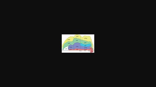
Retrosplenial cortex (RSC) has unique problems for human neuroimaging studies as its divisions are small, at the lower end of functional scanner spatial resolution, and it is buried in the callosal sulcus. The present study sought to define the cytoarchitecture of RSC in human and monkey brains along its entire anteroposterior extent. The results show anterior extensions, a newly defined dichotomy of area 30, a new area p30, and an area p29v in monkey that differentiates into three divisions in human. Accordingly, anterior (a), intermediate (i), and posterior (p) divisions of areas 29l, 29m, 30l, and 30m were identified. Posterior area 29 has higher neuron packing in the granular layer than anterior and intermediate divisions of area 29. A newly detected dysgranular area p30 has larger neurons in layers II–IIIab than a30 and i30 and with substantially higher NFP expression in layer IIIab of posterior areas than areas a30 and i30. Medial area 30 has larger pyramids and higher NFP expression in all layers than area 30l. The new area p30 was seen between areas p29m and p30I in both species. Finally, a ventral area p29v is present in monkeys. This latter area appears to differentiate into three divisions in human with the most extensive granular layer adjacent to layer I in p29vm and p29vl. Functional imaging has identified pRSC as part of a cognitive map which is engaged in spatial navigation and localization of personally relevant objects.
期刊介绍:
Established in 1891, JCN is the oldest continually published basic neuroscience journal. Historically, as the name suggests, the journal focused on a comparison among species to uncover the intricacies of how the brain functions. In modern times, this research is called systems neuroscience where animal models are used to mimic core cognitive processes with the ultimate goal of understanding neural circuits and connections that give rise to behavioral patterns and different neural states.
Research published in JCN covers all species from invertebrates to humans, and the reports inform the readers about the function and organization of nervous systems in species with an emphasis on the way that species adaptations inform about the function or organization of the nervous systems, rather than on their evolution per se.
JCN publishes primary research articles and critical commentaries and review-type articles offering expert insight in to cutting edge research in the field of systems neuroscience; a complete list of contribution types is given in the Author Guidelines. For primary research contributions, only full-length investigative reports are desired; the journal does not accept short communications.

 求助内容:
求助内容: 应助结果提醒方式:


