Gregory A. Lewbart, Giuliano Colosimo, Christopher Gaudette, Tatiane T. Negrão Watanabe, Joshua Parker, Christian Sevilla, Glenn P. Gerber, Gabriele Gentile
{"title":"当粉红色是一个问题:比较肉眼和显微镜下的皮肤结构分析揭示了Galápagos粉红地鬣蜥(Conolophus marthae)皮肤颜色的组织学基础","authors":"Gregory A. Lewbart, Giuliano Colosimo, Christopher Gaudette, Tatiane T. Negrão Watanabe, Joshua Parker, Christian Sevilla, Glenn P. Gerber, Gabriele Gentile","doi":"10.1111/azo.12488","DOIUrl":null,"url":null,"abstract":"<p>One of the rarest and most unusual iguanas on the planet is the Galápagos pink land iguana (<i>Conolophus marthae</i>). There have been a number of hypotheses on the source of their pink coloration, including that the colour is from blood and a relative lack of dermal pigmentation. We obtained full thickness skin biopsies of three species and compared tissue from darkly pigmented areas and lightly pigmented surfaces. “Pink” areas of pink iguanas are devoid of pigment cells (e.g. melanophores) and the dermal tissue is rich with aggregates of confluent capillaries. This was in sharp contrast to the minimally vascular (only capillaries were observed) dermal areas of the marine and yellow iguanas. The dermal stratum laxum of every biopsy site contained melanophores except for the pink skin of pink iguanas. Interestingly, marine iguanas have a much thicker epidermal stratum germinativum/granulosum, between 2 and 10 cells thick depending on location, compared to the thinner epidermal stratum germinativum/granulosum of land iguanas (one to three cells thick with most areas possessing just one or two cell layers). These microscopic differences might reflect differences in habitat and ecology of the three species.</p>","PeriodicalId":50945,"journal":{"name":"Acta Zoologica","volume":"105 4","pages":"514-523"},"PeriodicalIF":1.1000,"publicationDate":"2023-11-23","publicationTypes":"Journal Article","fieldsOfStudy":null,"isOpenAccess":false,"openAccessPdf":"https://onlinelibrary.wiley.com/doi/epdf/10.1111/azo.12488","citationCount":"0","resultStr":"{\"title\":\"When pink is a question: Comparative gross and microscopic skin structure analyses reveal the histological basis of skin colour in Galápagos pink land iguanas (Conolophus marthae)\",\"authors\":\"Gregory A. Lewbart, Giuliano Colosimo, Christopher Gaudette, Tatiane T. Negrão Watanabe, Joshua Parker, Christian Sevilla, Glenn P. Gerber, Gabriele Gentile\",\"doi\":\"10.1111/azo.12488\",\"DOIUrl\":null,\"url\":null,\"abstract\":\"<p>One of the rarest and most unusual iguanas on the planet is the Galápagos pink land iguana (<i>Conolophus marthae</i>). There have been a number of hypotheses on the source of their pink coloration, including that the colour is from blood and a relative lack of dermal pigmentation. We obtained full thickness skin biopsies of three species and compared tissue from darkly pigmented areas and lightly pigmented surfaces. “Pink” areas of pink iguanas are devoid of pigment cells (e.g. melanophores) and the dermal tissue is rich with aggregates of confluent capillaries. This was in sharp contrast to the minimally vascular (only capillaries were observed) dermal areas of the marine and yellow iguanas. The dermal stratum laxum of every biopsy site contained melanophores except for the pink skin of pink iguanas. Interestingly, marine iguanas have a much thicker epidermal stratum germinativum/granulosum, between 2 and 10 cells thick depending on location, compared to the thinner epidermal stratum germinativum/granulosum of land iguanas (one to three cells thick with most areas possessing just one or two cell layers). These microscopic differences might reflect differences in habitat and ecology of the three species.</p>\",\"PeriodicalId\":50945,\"journal\":{\"name\":\"Acta Zoologica\",\"volume\":\"105 4\",\"pages\":\"514-523\"},\"PeriodicalIF\":1.1000,\"publicationDate\":\"2023-11-23\",\"publicationTypes\":\"Journal Article\",\"fieldsOfStudy\":null,\"isOpenAccess\":false,\"openAccessPdf\":\"https://onlinelibrary.wiley.com/doi/epdf/10.1111/azo.12488\",\"citationCount\":\"0\",\"resultStr\":null,\"platform\":\"Semanticscholar\",\"paperid\":null,\"PeriodicalName\":\"Acta Zoologica\",\"FirstCategoryId\":\"99\",\"ListUrlMain\":\"https://onlinelibrary.wiley.com/doi/10.1111/azo.12488\",\"RegionNum\":4,\"RegionCategory\":\"生物学\",\"ArticlePicture\":[],\"TitleCN\":null,\"AbstractTextCN\":null,\"PMCID\":null,\"EPubDate\":\"\",\"PubModel\":\"\",\"JCR\":\"Q4\",\"JCRName\":\"ANATOMY & MORPHOLOGY\",\"Score\":null,\"Total\":0}","platform":"Semanticscholar","paperid":null,"PeriodicalName":"Acta Zoologica","FirstCategoryId":"99","ListUrlMain":"https://onlinelibrary.wiley.com/doi/10.1111/azo.12488","RegionNum":4,"RegionCategory":"生物学","ArticlePicture":[],"TitleCN":null,"AbstractTextCN":null,"PMCID":null,"EPubDate":"","PubModel":"","JCR":"Q4","JCRName":"ANATOMY & MORPHOLOGY","Score":null,"Total":0}
When pink is a question: Comparative gross and microscopic skin structure analyses reveal the histological basis of skin colour in Galápagos pink land iguanas (Conolophus marthae)
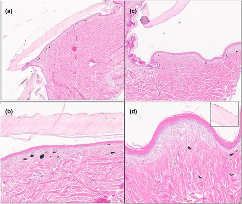
One of the rarest and most unusual iguanas on the planet is the Galápagos pink land iguana (Conolophus marthae). There have been a number of hypotheses on the source of their pink coloration, including that the colour is from blood and a relative lack of dermal pigmentation. We obtained full thickness skin biopsies of three species and compared tissue from darkly pigmented areas and lightly pigmented surfaces. “Pink” areas of pink iguanas are devoid of pigment cells (e.g. melanophores) and the dermal tissue is rich with aggregates of confluent capillaries. This was in sharp contrast to the minimally vascular (only capillaries were observed) dermal areas of the marine and yellow iguanas. The dermal stratum laxum of every biopsy site contained melanophores except for the pink skin of pink iguanas. Interestingly, marine iguanas have a much thicker epidermal stratum germinativum/granulosum, between 2 and 10 cells thick depending on location, compared to the thinner epidermal stratum germinativum/granulosum of land iguanas (one to three cells thick with most areas possessing just one or two cell layers). These microscopic differences might reflect differences in habitat and ecology of the three species.
期刊介绍:
Published regularly since 1920, Acta Zoologica has retained its position as one of the world''s leading journals in the field of animal organization, development, structure and function. Each issue publishes original research of interest to zoologists and physiologists worldwide, in the field of animal structure (from the cellular to the organismic level) and development with emphasis on functional, comparative and phylogenetic aspects. Occasional review articles are also published, as well as book reviews.
 求助内容:
求助内容: 应助结果提醒方式:


