Roman Taday, Erik Schiffner, Sebastian Viktor Gehrmann, Lena Marie Wilms, Robert Alexander Kaufmann, Joachim Windolf, David Latz
{"title":"利用便携式3D扫描仪在骨科和创伤外科中建立小腿和脚踝围手术期体积评估的感兴趣区域-一项试点研究。","authors":"Roman Taday, Erik Schiffner, Sebastian Viktor Gehrmann, Lena Marie Wilms, Robert Alexander Kaufmann, Joachim Windolf, David Latz","doi":"10.1186/s13047-023-00684-2","DOIUrl":null,"url":null,"abstract":"<p><strong>Background: </strong>Soft tissue swelling assessment benefits from a reproducible and easy to use measurement method. Monitoring of the injured lower extremity is of clinical import during staged soft tissue management. Portable 3D scanners offer a novel and precise option to quantify and contrast the shapes and volumes of the injured and contralateral uninjured limbs. This study determined three regions of interest (ROI) within the lower extremity (lower leg, ankle and foot), that can be used to evaluate 3D volumetric assessment for staged soft tissue management in orthopedic and trauma surgery.</p><p><strong>Methods: </strong>Twelve healthy volunteers (24 legs) were included in this cohort study. Scans of all three ROI were recorded with a portable 3D scanner (Artec, 3D scanner EVA) and compared between the right and left leg using the software Artec Studio (Arctec Group, Luxemburg).</p><p><strong>Results: </strong>Mean volume of the right leg was 1926.64 ± 308.84 ml (mean ± SD). ROI: lower leg: 931.86 ± 236.15 ml; ankle: 201.56 ± 27.88 ml; foot: 793.21 ± 112.28 ml. Mean volume of the left leg was 1937.73 ± 329.51 ml. ROI: lower leg: 933.59 ± 251.12 ml; ankle: 201.53 ± 25.54 ml; foot: 802.62 ± 124.83 ml. There was no significant difference of the overall volume between right and left leg (p > 0.05; overall volume: △ difference: 29.5 ± 7.29 ml, p = 0.8; lower leg: △ difference: 21.5 ± 6.39 ml, p = 0.8; ankle: △ difference: 5.3 ± 2.11 ml, p = 0.4; △ difference: 16.33 ± 4.45 ml, p = 0.8.</p><p><strong>Conclusion: </strong>This pilot study defines three regions of interest of the lower leg and demonstrates no difference between the right and left side. Based on these ROI, further studies are needed to evaluate the clinical applicability of the scanner.</p>","PeriodicalId":49164,"journal":{"name":"Journal of Foot and Ankle Research","volume":"16 1","pages":"87"},"PeriodicalIF":2.2000,"publicationDate":"2023-12-05","publicationTypes":"Journal Article","fieldsOfStudy":null,"isOpenAccess":false,"openAccessPdf":"https://www.ncbi.nlm.nih.gov/pmc/articles/PMC10696714/pdf/","citationCount":"0","resultStr":"{\"title\":\"Establishing regions of interest of the lower leg and ankle for perioperative volumetric assessment with a portable 3D scanner in orthopedic and trauma surgery - a pilot study.\",\"authors\":\"Roman Taday, Erik Schiffner, Sebastian Viktor Gehrmann, Lena Marie Wilms, Robert Alexander Kaufmann, Joachim Windolf, David Latz\",\"doi\":\"10.1186/s13047-023-00684-2\",\"DOIUrl\":null,\"url\":null,\"abstract\":\"<p><strong>Background: </strong>Soft tissue swelling assessment benefits from a reproducible and easy to use measurement method. Monitoring of the injured lower extremity is of clinical import during staged soft tissue management. Portable 3D scanners offer a novel and precise option to quantify and contrast the shapes and volumes of the injured and contralateral uninjured limbs. This study determined three regions of interest (ROI) within the lower extremity (lower leg, ankle and foot), that can be used to evaluate 3D volumetric assessment for staged soft tissue management in orthopedic and trauma surgery.</p><p><strong>Methods: </strong>Twelve healthy volunteers (24 legs) were included in this cohort study. Scans of all three ROI were recorded with a portable 3D scanner (Artec, 3D scanner EVA) and compared between the right and left leg using the software Artec Studio (Arctec Group, Luxemburg).</p><p><strong>Results: </strong>Mean volume of the right leg was 1926.64 ± 308.84 ml (mean ± SD). ROI: lower leg: 931.86 ± 236.15 ml; ankle: 201.56 ± 27.88 ml; foot: 793.21 ± 112.28 ml. Mean volume of the left leg was 1937.73 ± 329.51 ml. ROI: lower leg: 933.59 ± 251.12 ml; ankle: 201.53 ± 25.54 ml; foot: 802.62 ± 124.83 ml. There was no significant difference of the overall volume between right and left leg (p > 0.05; overall volume: △ difference: 29.5 ± 7.29 ml, p = 0.8; lower leg: △ difference: 21.5 ± 6.39 ml, p = 0.8; ankle: △ difference: 5.3 ± 2.11 ml, p = 0.4; △ difference: 16.33 ± 4.45 ml, p = 0.8.</p><p><strong>Conclusion: </strong>This pilot study defines three regions of interest of the lower leg and demonstrates no difference between the right and left side. Based on these ROI, further studies are needed to evaluate the clinical applicability of the scanner.</p>\",\"PeriodicalId\":49164,\"journal\":{\"name\":\"Journal of Foot and Ankle Research\",\"volume\":\"16 1\",\"pages\":\"87\"},\"PeriodicalIF\":2.2000,\"publicationDate\":\"2023-12-05\",\"publicationTypes\":\"Journal Article\",\"fieldsOfStudy\":null,\"isOpenAccess\":false,\"openAccessPdf\":\"https://www.ncbi.nlm.nih.gov/pmc/articles/PMC10696714/pdf/\",\"citationCount\":\"0\",\"resultStr\":null,\"platform\":\"Semanticscholar\",\"paperid\":null,\"PeriodicalName\":\"Journal of Foot and Ankle Research\",\"FirstCategoryId\":\"3\",\"ListUrlMain\":\"https://doi.org/10.1186/s13047-023-00684-2\",\"RegionNum\":3,\"RegionCategory\":\"医学\",\"ArticlePicture\":[],\"TitleCN\":null,\"AbstractTextCN\":null,\"PMCID\":null,\"EPubDate\":\"\",\"PubModel\":\"\",\"JCR\":\"Q1\",\"JCRName\":\"ORTHOPEDICS\",\"Score\":null,\"Total\":0}","platform":"Semanticscholar","paperid":null,"PeriodicalName":"Journal of Foot and Ankle Research","FirstCategoryId":"3","ListUrlMain":"https://doi.org/10.1186/s13047-023-00684-2","RegionNum":3,"RegionCategory":"医学","ArticlePicture":[],"TitleCN":null,"AbstractTextCN":null,"PMCID":null,"EPubDate":"","PubModel":"","JCR":"Q1","JCRName":"ORTHOPEDICS","Score":null,"Total":0}
引用次数: 0
摘要
背景:软组织肿胀评估得益于一种可重复且易于使用的测量方法。监测损伤的下肢是临床上重要的阶段性软组织处理。便携式3D扫描仪提供了一种新颖而精确的选择,可以量化和对比受伤和对侧未受伤肢体的形状和体积。本研究确定了下肢(小腿、脚踝和足)内的三个感兴趣区域(ROI),可用于评估骨科和创伤手术中分阶段软组织管理的3D体积评估。方法:12名健康志愿者(24条腿)加入队列研究。使用便携式3D扫描仪(Artec, 3D扫描仪EVA)记录所有三个ROI的扫描结果,并使用Artec Studio软件(Arctec Group, Luxemburg)在右腿和左腿之间进行比较。结果:右腿平均容积为1926.64±308.84 ml (Mean±SD)。ROI:小腿:931.86±236.15 ml;踝关节:201.56±27.88 ml;足部:793.21±112.28 ml,左腿平均容积:1937.73±329.51 ml,下肢平均容积:933.59±251.12 ml;踝关节:201.53±25.54 ml;足部:802.62±124.83 ml,左右两腿总容积差异无统计学意义(p > 0.05);总体容积:△差值:29.5±7.29 ml, p = 0.8;小腿:△差异:21.5±6.39 ml, p = 0.8;踝关节:△差异:5.3±2.11 ml, p = 0.4;△差异:16.33±4.45 ml, p = 0.8。结论:这项初步研究确定了下肢的三个感兴趣区域,并表明左右侧没有差异。基于这些ROI,需要进一步的研究来评估扫描仪的临床适用性。
Establishing regions of interest of the lower leg and ankle for perioperative volumetric assessment with a portable 3D scanner in orthopedic and trauma surgery - a pilot study.
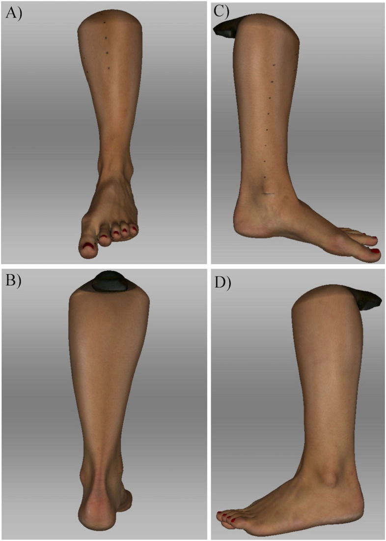
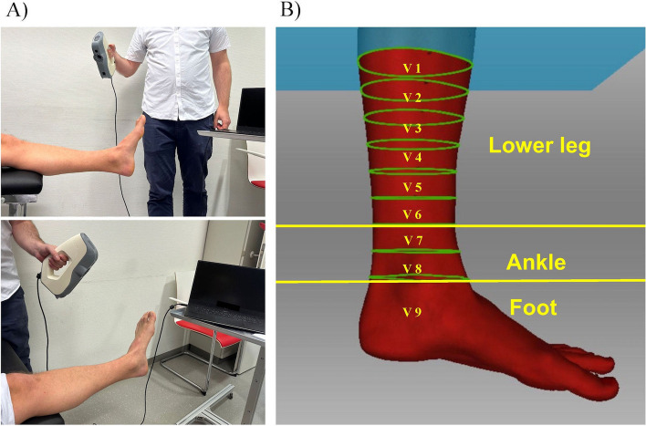
Background: Soft tissue swelling assessment benefits from a reproducible and easy to use measurement method. Monitoring of the injured lower extremity is of clinical import during staged soft tissue management. Portable 3D scanners offer a novel and precise option to quantify and contrast the shapes and volumes of the injured and contralateral uninjured limbs. This study determined three regions of interest (ROI) within the lower extremity (lower leg, ankle and foot), that can be used to evaluate 3D volumetric assessment for staged soft tissue management in orthopedic and trauma surgery.
Methods: Twelve healthy volunteers (24 legs) were included in this cohort study. Scans of all three ROI were recorded with a portable 3D scanner (Artec, 3D scanner EVA) and compared between the right and left leg using the software Artec Studio (Arctec Group, Luxemburg).
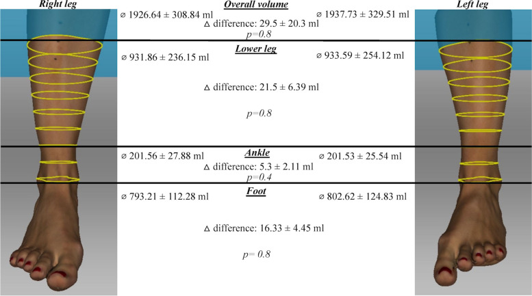
Results: Mean volume of the right leg was 1926.64 ± 308.84 ml (mean ± SD). ROI: lower leg: 931.86 ± 236.15 ml; ankle: 201.56 ± 27.88 ml; foot: 793.21 ± 112.28 ml. Mean volume of the left leg was 1937.73 ± 329.51 ml. ROI: lower leg: 933.59 ± 251.12 ml; ankle: 201.53 ± 25.54 ml; foot: 802.62 ± 124.83 ml. There was no significant difference of the overall volume between right and left leg (p > 0.05; overall volume: △ difference: 29.5 ± 7.29 ml, p = 0.8; lower leg: △ difference: 21.5 ± 6.39 ml, p = 0.8; ankle: △ difference: 5.3 ± 2.11 ml, p = 0.4; △ difference: 16.33 ± 4.45 ml, p = 0.8.
Conclusion: This pilot study defines three regions of interest of the lower leg and demonstrates no difference between the right and left side. Based on these ROI, further studies are needed to evaluate the clinical applicability of the scanner.
期刊介绍:
Journal of Foot and Ankle Research, the official journal of the Australian Podiatry Association and The College of Podiatry (UK), is an open access journal that encompasses all aspects of policy, organisation, delivery and clinical practice related to the assessment, diagnosis, prevention and management of foot and ankle disorders.
Journal of Foot and Ankle Research covers a wide range of clinical subject areas, including diabetology, paediatrics, sports medicine, gerontology and geriatrics, foot surgery, physical therapy, dermatology, wound management, radiology, biomechanics and bioengineering, orthotics and prosthetics, as well the broad areas of epidemiology, policy, organisation and delivery of services related to foot and ankle care.
The journal encourages submissions from all health professionals who manage lower limb conditions, including podiatrists, nurses, physical therapists and physiotherapists, orthopaedists, manual therapists, medical specialists and general medical practitioners, as well as health service researchers concerned with foot and ankle care.
The Australian Podiatry Association and the College of Podiatry (UK) have reserve funds to cover the article-processing charge for manuscripts submitted by its members. Society members can email the appropriate contact at Australian Podiatry Association or The College of Podiatry to obtain the corresponding code to enter on submission.
 求助内容:
求助内容: 应助结果提醒方式:


