Arvind Kumar, Tushar Kalonia, Akanksha Gupta, Ravi Hari Phulware
{"title":"胆囊腺癌皮肤转移。","authors":"Arvind Kumar, Tushar Kalonia, Akanksha Gupta, Ravi Hari Phulware","doi":"10.4322/acr.2023.458","DOIUrl":null,"url":null,"abstract":"moderate cytoplasm. A few cells showed cytoplasmic clearing. The peritumoral desmoplastic reaction was also identified with intratumorally brisk mitotic figures. Focal areas of necrosis with acute and chronic inflammatory infiltrate were also identified. Immunohistochemistry revealed tumor cells to be diffusely positive for cytokeratin 7 (CK7) and CK19 (Figure 1C and 1D) and negative for CK20, thyroid transcription factor (TTF-1), Gross Cystic Disease Fluid Protein-15 (GCDFP-15), mammaglobin, Hepatocyte Paraffin 1 (Hep Par1), Paired box gene 8 (PAX-8) and Wilms tumor gene 1 (WT1). An abdominal computed tomography scan on imaging workup revealed a gallbladder mass with multiple liver lesions and brain metastasis. Based on immunohistochemistry and imaging findings, the diagnosis was metastatic gall bladder carcinoma to the scalp.","PeriodicalId":53117,"journal":{"name":"Autopsy and Case Reports","volume":"13 ","pages":"e2023458"},"PeriodicalIF":0.0000,"publicationDate":"2023-11-27","publicationTypes":"Journal Article","fieldsOfStudy":null,"isOpenAccess":false,"openAccessPdf":"https://www.ncbi.nlm.nih.gov/pmc/articles/PMC10688260/pdf/","citationCount":"0","resultStr":"{\"title\":\"Gallbladder adenocarcinoma skin metastasis.\",\"authors\":\"Arvind Kumar, Tushar Kalonia, Akanksha Gupta, Ravi Hari Phulware\",\"doi\":\"10.4322/acr.2023.458\",\"DOIUrl\":null,\"url\":null,\"abstract\":\"moderate cytoplasm. A few cells showed cytoplasmic clearing. The peritumoral desmoplastic reaction was also identified with intratumorally brisk mitotic figures. Focal areas of necrosis with acute and chronic inflammatory infiltrate were also identified. Immunohistochemistry revealed tumor cells to be diffusely positive for cytokeratin 7 (CK7) and CK19 (Figure 1C and 1D) and negative for CK20, thyroid transcription factor (TTF-1), Gross Cystic Disease Fluid Protein-15 (GCDFP-15), mammaglobin, Hepatocyte Paraffin 1 (Hep Par1), Paired box gene 8 (PAX-8) and Wilms tumor gene 1 (WT1). An abdominal computed tomography scan on imaging workup revealed a gallbladder mass with multiple liver lesions and brain metastasis. Based on immunohistochemistry and imaging findings, the diagnosis was metastatic gall bladder carcinoma to the scalp.\",\"PeriodicalId\":53117,\"journal\":{\"name\":\"Autopsy and Case Reports\",\"volume\":\"13 \",\"pages\":\"e2023458\"},\"PeriodicalIF\":0.0000,\"publicationDate\":\"2023-11-27\",\"publicationTypes\":\"Journal Article\",\"fieldsOfStudy\":null,\"isOpenAccess\":false,\"openAccessPdf\":\"https://www.ncbi.nlm.nih.gov/pmc/articles/PMC10688260/pdf/\",\"citationCount\":\"0\",\"resultStr\":null,\"platform\":\"Semanticscholar\",\"paperid\":null,\"PeriodicalName\":\"Autopsy and Case Reports\",\"FirstCategoryId\":\"1085\",\"ListUrlMain\":\"https://doi.org/10.4322/acr.2023.458\",\"RegionNum\":0,\"RegionCategory\":null,\"ArticlePicture\":[],\"TitleCN\":null,\"AbstractTextCN\":null,\"PMCID\":null,\"EPubDate\":\"2023/1/1 0:00:00\",\"PubModel\":\"eCollection\",\"JCR\":\"Q4\",\"JCRName\":\"Medicine\",\"Score\":null,\"Total\":0}","platform":"Semanticscholar","paperid":null,"PeriodicalName":"Autopsy and Case Reports","FirstCategoryId":"1085","ListUrlMain":"https://doi.org/10.4322/acr.2023.458","RegionNum":0,"RegionCategory":null,"ArticlePicture":[],"TitleCN":null,"AbstractTextCN":null,"PMCID":null,"EPubDate":"2023/1/1 0:00:00","PubModel":"eCollection","JCR":"Q4","JCRName":"Medicine","Score":null,"Total":0}
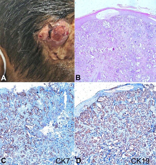
moderate cytoplasm. A few cells showed cytoplasmic clearing. The peritumoral desmoplastic reaction was also identified with intratumorally brisk mitotic figures. Focal areas of necrosis with acute and chronic inflammatory infiltrate were also identified. Immunohistochemistry revealed tumor cells to be diffusely positive for cytokeratin 7 (CK7) and CK19 (Figure 1C and 1D) and negative for CK20, thyroid transcription factor (TTF-1), Gross Cystic Disease Fluid Protein-15 (GCDFP-15), mammaglobin, Hepatocyte Paraffin 1 (Hep Par1), Paired box gene 8 (PAX-8) and Wilms tumor gene 1 (WT1). An abdominal computed tomography scan on imaging workup revealed a gallbladder mass with multiple liver lesions and brain metastasis. Based on immunohistochemistry and imaging findings, the diagnosis was metastatic gall bladder carcinoma to the scalp.