Cristina Velasquillo, Yaaziel Melgarejo-Ramírez, Julieta García-López, Claudia Gutiérrez-Gómez, Hugo Lecona, Maykel González-Torres, José Iván Sánchez-Betancourt, Clemente Ibarra, Sang Jin Lee, James J Yoo
{"title":"剩余的小体组织作为3D生物打印弹性软骨组织结构的来源,可能用于外科小体重建。","authors":"Cristina Velasquillo, Yaaziel Melgarejo-Ramírez, Julieta García-López, Claudia Gutiérrez-Gómez, Hugo Lecona, Maykel González-Torres, José Iván Sánchez-Betancourt, Clemente Ibarra, Sang Jin Lee, James J Yoo","doi":"10.1007/s10561-023-10118-9","DOIUrl":null,"url":null,"abstract":"<p><p>The absence of ears in children is a global problem. An implant made of costal cartilage is the standard procedure for ear reconstruction; however, side effects such as pneumothorax, loss of thoracic cage shape, and respiratory complications have been documented. Three-dimensional (3D) printing allows the generation of biocompatible scaffolds that mimic the shape, mechanical strength, and architecture of the native extracellular matrix necessary to promote new elastic cartilage formation. We report the potential use of a 3D-bioprinted poly-ε-caprolactone (3D-PCL) auricle-shaped framework seeded with remaining human microtia chondrocytes for the development of elastic cartilage for autologous microtia ear reconstruction. An in vivo assay of the neo-tissue formed revealed the generation of a 3D pinna-shaped neo-tissue, and confirmed the formation of elastic cartilage by the presence of type II collagen and elastin with histological features and a protein composition consistent with normal elastic cartilage. According to our results, a combination of 3D-PCL auricle frameworks and autologous microtia remnant tissue generates a suitable pinna structure for autologous ear reconstruction.</p>","PeriodicalId":9723,"journal":{"name":"Cell and Tissue Banking","volume":" ","pages":"571-582"},"PeriodicalIF":1.4000,"publicationDate":"2024-06-01","publicationTypes":"Journal Article","fieldsOfStudy":null,"isOpenAccess":false,"openAccessPdf":"","citationCount":"0","resultStr":"{\"title\":\"Remaining microtia tissue as a source for 3D bioprinted elastic cartilage tissue constructs, potential use for surgical microtia reconstruction.\",\"authors\":\"Cristina Velasquillo, Yaaziel Melgarejo-Ramírez, Julieta García-López, Claudia Gutiérrez-Gómez, Hugo Lecona, Maykel González-Torres, José Iván Sánchez-Betancourt, Clemente Ibarra, Sang Jin Lee, James J Yoo\",\"doi\":\"10.1007/s10561-023-10118-9\",\"DOIUrl\":null,\"url\":null,\"abstract\":\"<p><p>The absence of ears in children is a global problem. An implant made of costal cartilage is the standard procedure for ear reconstruction; however, side effects such as pneumothorax, loss of thoracic cage shape, and respiratory complications have been documented. Three-dimensional (3D) printing allows the generation of biocompatible scaffolds that mimic the shape, mechanical strength, and architecture of the native extracellular matrix necessary to promote new elastic cartilage formation. We report the potential use of a 3D-bioprinted poly-ε-caprolactone (3D-PCL) auricle-shaped framework seeded with remaining human microtia chondrocytes for the development of elastic cartilage for autologous microtia ear reconstruction. An in vivo assay of the neo-tissue formed revealed the generation of a 3D pinna-shaped neo-tissue, and confirmed the formation of elastic cartilage by the presence of type II collagen and elastin with histological features and a protein composition consistent with normal elastic cartilage. According to our results, a combination of 3D-PCL auricle frameworks and autologous microtia remnant tissue generates a suitable pinna structure for autologous ear reconstruction.</p>\",\"PeriodicalId\":9723,\"journal\":{\"name\":\"Cell and Tissue Banking\",\"volume\":\" \",\"pages\":\"571-582\"},\"PeriodicalIF\":1.4000,\"publicationDate\":\"2024-06-01\",\"publicationTypes\":\"Journal Article\",\"fieldsOfStudy\":null,\"isOpenAccess\":false,\"openAccessPdf\":\"\",\"citationCount\":\"0\",\"resultStr\":null,\"platform\":\"Semanticscholar\",\"paperid\":null,\"PeriodicalName\":\"Cell and Tissue Banking\",\"FirstCategoryId\":\"5\",\"ListUrlMain\":\"https://doi.org/10.1007/s10561-023-10118-9\",\"RegionNum\":4,\"RegionCategory\":\"医学\",\"ArticlePicture\":[],\"TitleCN\":null,\"AbstractTextCN\":null,\"PMCID\":null,\"EPubDate\":\"2023/12/1 0:00:00\",\"PubModel\":\"Epub\",\"JCR\":\"Q4\",\"JCRName\":\"CELL BIOLOGY\",\"Score\":null,\"Total\":0}","platform":"Semanticscholar","paperid":null,"PeriodicalName":"Cell and Tissue Banking","FirstCategoryId":"5","ListUrlMain":"https://doi.org/10.1007/s10561-023-10118-9","RegionNum":4,"RegionCategory":"医学","ArticlePicture":[],"TitleCN":null,"AbstractTextCN":null,"PMCID":null,"EPubDate":"2023/12/1 0:00:00","PubModel":"Epub","JCR":"Q4","JCRName":"CELL BIOLOGY","Score":null,"Total":0}
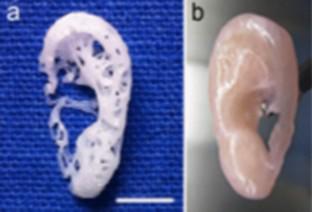
Remaining microtia tissue as a source for 3D bioprinted elastic cartilage tissue constructs, potential use for surgical microtia reconstruction.
The absence of ears in children is a global problem. An implant made of costal cartilage is the standard procedure for ear reconstruction; however, side effects such as pneumothorax, loss of thoracic cage shape, and respiratory complications have been documented. Three-dimensional (3D) printing allows the generation of biocompatible scaffolds that mimic the shape, mechanical strength, and architecture of the native extracellular matrix necessary to promote new elastic cartilage formation. We report the potential use of a 3D-bioprinted poly-ε-caprolactone (3D-PCL) auricle-shaped framework seeded with remaining human microtia chondrocytes for the development of elastic cartilage for autologous microtia ear reconstruction. An in vivo assay of the neo-tissue formed revealed the generation of a 3D pinna-shaped neo-tissue, and confirmed the formation of elastic cartilage by the presence of type II collagen and elastin with histological features and a protein composition consistent with normal elastic cartilage. According to our results, a combination of 3D-PCL auricle frameworks and autologous microtia remnant tissue generates a suitable pinna structure for autologous ear reconstruction.
期刊介绍:
Cell and Tissue Banking provides a forum for disseminating information to scientists and clinicians involved in the banking and transplantation of cells and tissues. Cell and Tissue Banking is an international, peer-reviewed journal that publishes original papers in the following areas:
basic research concerning general aspects of tissue banking such as quality assurance and control of banked cells/tissues, effects of preservation and sterilisation methods on cells/tissues, biotechnology, etc.; clinical applications of banked cells/tissues; standards of practice in procurement, processing, storage and distribution of cells/tissues; ethical issues; medico-legal issues.
 求助内容:
求助内容: 应助结果提醒方式:


