Nishama De Silva Mohotti, Hiroko Kobayashi, Jenna M Williams, Rashmi Binjawadagi, Michel P Evertsen, Ethan G Christ, Meredith D Hartley
{"title":"脂质组学分析揭示脑和脊髓再髓鞘形成程度的差异。","authors":"Nishama De Silva Mohotti, Hiroko Kobayashi, Jenna M Williams, Rashmi Binjawadagi, Michel P Evertsen, Ethan G Christ, Meredith D Hartley","doi":"10.1021/acs.jproteome.3c00443","DOIUrl":null,"url":null,"abstract":"<p><p>During demyelination, lipid-rich myelin debris is released in the central nervous system (CNS) and must be phagocytosed and processed before new myelin can form. Although myelin comprises over 70% lipids, relatively little is known about how the CNS lipidome changes during demyelination and remyelination. In this study, we obtained a longitudinal lipidomic profile of the brain, spinal cord, and serum using a genetic mouse model of demyelination, known as <i>Plp1</i>-iCKO-<i>Myrf</i>. The mass spectrometry data is available at the Metabolomics Workbench, where it has been assigned Study ID ST002958. This model has distinct phases of demyelination and remyelination over the course of 24 weeks, in which loss of motor function peaks during demyelination. Using principal component analysis (PCA) and volcano plots, we have demonstrated that the brain and spinal cord have different remyelination capabilities and that this is reflected in different lipidomic profiles over time. We observed that plasmalogens (ether-linked phosphatidylserine and ether-linked phosphatidylcholine) were elevated specifically during the early stages of active demyelination. In addition, we identified lipids in the brain that were altered when mice were treated with a remyelinating drug, which may be CNS biomarkers of remyelination. The results of this study provide new insights into how the lipidome changes in response to demyelination, which will enable future studies to elucidate mechanisms of lipid regulation during demyelination and remyelination.</p>","PeriodicalId":48,"journal":{"name":"Journal of Proteome Research","volume":" ","pages":"2830-2844"},"PeriodicalIF":3.6000,"publicationDate":"2024-08-02","publicationTypes":"Journal Article","fieldsOfStudy":null,"isOpenAccess":false,"openAccessPdf":"https://www.ncbi.nlm.nih.gov/pmc/articles/PMC11133230/pdf/","citationCount":"0","resultStr":"{\"title\":\"Lipidomic Analysis Reveals Differences in the Extent of Remyelination in the Brain and Spinal Cord.\",\"authors\":\"Nishama De Silva Mohotti, Hiroko Kobayashi, Jenna M Williams, Rashmi Binjawadagi, Michel P Evertsen, Ethan G Christ, Meredith D Hartley\",\"doi\":\"10.1021/acs.jproteome.3c00443\",\"DOIUrl\":null,\"url\":null,\"abstract\":\"<p><p>During demyelination, lipid-rich myelin debris is released in the central nervous system (CNS) and must be phagocytosed and processed before new myelin can form. Although myelin comprises over 70% lipids, relatively little is known about how the CNS lipidome changes during demyelination and remyelination. In this study, we obtained a longitudinal lipidomic profile of the brain, spinal cord, and serum using a genetic mouse model of demyelination, known as <i>Plp1</i>-iCKO-<i>Myrf</i>. The mass spectrometry data is available at the Metabolomics Workbench, where it has been assigned Study ID ST002958. This model has distinct phases of demyelination and remyelination over the course of 24 weeks, in which loss of motor function peaks during demyelination. Using principal component analysis (PCA) and volcano plots, we have demonstrated that the brain and spinal cord have different remyelination capabilities and that this is reflected in different lipidomic profiles over time. We observed that plasmalogens (ether-linked phosphatidylserine and ether-linked phosphatidylcholine) were elevated specifically during the early stages of active demyelination. In addition, we identified lipids in the brain that were altered when mice were treated with a remyelinating drug, which may be CNS biomarkers of remyelination. The results of this study provide new insights into how the lipidome changes in response to demyelination, which will enable future studies to elucidate mechanisms of lipid regulation during demyelination and remyelination.</p>\",\"PeriodicalId\":48,\"journal\":{\"name\":\"Journal of Proteome Research\",\"volume\":\" \",\"pages\":\"2830-2844\"},\"PeriodicalIF\":3.6000,\"publicationDate\":\"2024-08-02\",\"publicationTypes\":\"Journal Article\",\"fieldsOfStudy\":null,\"isOpenAccess\":false,\"openAccessPdf\":\"https://www.ncbi.nlm.nih.gov/pmc/articles/PMC11133230/pdf/\",\"citationCount\":\"0\",\"resultStr\":null,\"platform\":\"Semanticscholar\",\"paperid\":null,\"PeriodicalName\":\"Journal of Proteome Research\",\"FirstCategoryId\":\"99\",\"ListUrlMain\":\"https://doi.org/10.1021/acs.jproteome.3c00443\",\"RegionNum\":2,\"RegionCategory\":\"生物学\",\"ArticlePicture\":[],\"TitleCN\":null,\"AbstractTextCN\":null,\"PMCID\":null,\"EPubDate\":\"2023/11/29 0:00:00\",\"PubModel\":\"Epub\",\"JCR\":\"Q1\",\"JCRName\":\"BIOCHEMICAL RESEARCH METHODS\",\"Score\":null,\"Total\":0}","platform":"Semanticscholar","paperid":null,"PeriodicalName":"Journal of Proteome Research","FirstCategoryId":"99","ListUrlMain":"https://doi.org/10.1021/acs.jproteome.3c00443","RegionNum":2,"RegionCategory":"生物学","ArticlePicture":[],"TitleCN":null,"AbstractTextCN":null,"PMCID":null,"EPubDate":"2023/11/29 0:00:00","PubModel":"Epub","JCR":"Q1","JCRName":"BIOCHEMICAL RESEARCH METHODS","Score":null,"Total":0}
Lipidomic Analysis Reveals Differences in the Extent of Remyelination in the Brain and Spinal Cord.
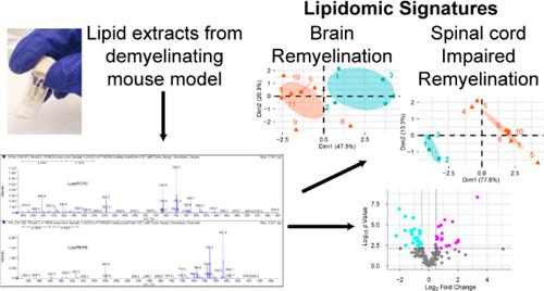
During demyelination, lipid-rich myelin debris is released in the central nervous system (CNS) and must be phagocytosed and processed before new myelin can form. Although myelin comprises over 70% lipids, relatively little is known about how the CNS lipidome changes during demyelination and remyelination. In this study, we obtained a longitudinal lipidomic profile of the brain, spinal cord, and serum using a genetic mouse model of demyelination, known as Plp1-iCKO-Myrf. The mass spectrometry data is available at the Metabolomics Workbench, where it has been assigned Study ID ST002958. This model has distinct phases of demyelination and remyelination over the course of 24 weeks, in which loss of motor function peaks during demyelination. Using principal component analysis (PCA) and volcano plots, we have demonstrated that the brain and spinal cord have different remyelination capabilities and that this is reflected in different lipidomic profiles over time. We observed that plasmalogens (ether-linked phosphatidylserine and ether-linked phosphatidylcholine) were elevated specifically during the early stages of active demyelination. In addition, we identified lipids in the brain that were altered when mice were treated with a remyelinating drug, which may be CNS biomarkers of remyelination. The results of this study provide new insights into how the lipidome changes in response to demyelination, which will enable future studies to elucidate mechanisms of lipid regulation during demyelination and remyelination.
期刊介绍:
Journal of Proteome Research publishes content encompassing all aspects of global protein analysis and function, including the dynamic aspects of genomics, spatio-temporal proteomics, metabonomics and metabolomics, clinical and agricultural proteomics, as well as advances in methodology including bioinformatics. The theme and emphasis is on a multidisciplinary approach to the life sciences through the synergy between the different types of "omics".
 求助内容:
求助内容: 应助结果提醒方式:


