May Sadik, Sally F. Barrington, Elin Trägårdh, Babak Saboury, Anne L. Nielsen, Annika L. Jakobsen, Jose L. L. Gongora, Jesus L. Urdaneta, Rajender Kumar, Lars Edenbrandt
{"title":"霍奇金淋巴瘤的代谢肿瘤体积-人工和人工智能分析的比较。","authors":"May Sadik, Sally F. Barrington, Elin Trägårdh, Babak Saboury, Anne L. Nielsen, Annika L. Jakobsen, Jose L. L. Gongora, Jesus L. Urdaneta, Rajender Kumar, Lars Edenbrandt","doi":"10.1111/cpf.12868","DOIUrl":null,"url":null,"abstract":"<div>\n \n \n <section>\n \n <h3> Aim</h3>\n \n <p>To compare total metabolic tumour volume (tMTV), calculated using two artificial intelligence (AI)-based tools, with manual segmentation by specialists as the reference.</p>\n </section>\n \n <section>\n \n <h3> Methods</h3>\n \n <p>Forty-eight consecutive Hodgkin lymphoma (HL) patients staged with [18F] fluorodeoxyglucose positron emission tomography/computed tomography were included. The median age was 35 years (range: 7–75), 46% female. The tMTV was automatically measured using the AI-based tools positron emission tomography assisted reporting system (PARS) (from Siemens) and RECOMIA (recomia.org) without any manual adjustments. A group of eight nuclear medicine specialists manually segmented lesions for tMTV calculations; each patient was independently segmented by two specialists.</p>\n </section>\n \n <section>\n \n <h3> Results</h3>\n \n <p>The median of the manual tMTV was 146 cm<sup>3</sup> (interquartile range [IQR]: 79–568 cm<sup>3</sup>) and the median difference between two tMTV values segmented by different specialists for the same patient was 26 cm<sup>3</sup> (IQR: 10–86 cm<sup>3</sup>). In 22 of the 48 patients, the manual tMTV value was closer to the RECOMIA tMTV value than to the manual tMTV value segmented by the second specialist. In 11 of the remaining 26 patients, the difference between the RECOMIA tMTV and the manual tMTV was small (<26 cm<sup>3</sup>, which was the median difference between two manual tMTV values from the same patient). The corresponding numbers for PARS were 18 and 10 patients, respectively.</p>\n </section>\n \n <section>\n \n <h3> Conclusion</h3>\n \n <p>The results of this study indicate that RECOMIA and Siemens PARS AI tools could be used without any major manual adjustments in 69% (33/48) and 58% (28/48) of HL patients, respectively. This demonstrates the feasibility of using AI tools to support physicians measuring tMTV for assessment of prognosis in clinical practice.</p>\n </section>\n </div>","PeriodicalId":10504,"journal":{"name":"Clinical Physiology and Functional Imaging","volume":"44 3","pages":"220-227"},"PeriodicalIF":1.3000,"publicationDate":"2023-11-27","publicationTypes":"Journal Article","fieldsOfStudy":null,"isOpenAccess":false,"openAccessPdf":"https://onlinelibrary.wiley.com/doi/epdf/10.1111/cpf.12868","citationCount":"0","resultStr":"{\"title\":\"Metabolic tumour volume in Hodgkin lymphoma—A comparison between manual and AI-based analysis\",\"authors\":\"May Sadik, Sally F. Barrington, Elin Trägårdh, Babak Saboury, Anne L. Nielsen, Annika L. Jakobsen, Jose L. L. Gongora, Jesus L. Urdaneta, Rajender Kumar, Lars Edenbrandt\",\"doi\":\"10.1111/cpf.12868\",\"DOIUrl\":null,\"url\":null,\"abstract\":\"<div>\\n \\n \\n <section>\\n \\n <h3> Aim</h3>\\n \\n <p>To compare total metabolic tumour volume (tMTV), calculated using two artificial intelligence (AI)-based tools, with manual segmentation by specialists as the reference.</p>\\n </section>\\n \\n <section>\\n \\n <h3> Methods</h3>\\n \\n <p>Forty-eight consecutive Hodgkin lymphoma (HL) patients staged with [18F] fluorodeoxyglucose positron emission tomography/computed tomography were included. The median age was 35 years (range: 7–75), 46% female. The tMTV was automatically measured using the AI-based tools positron emission tomography assisted reporting system (PARS) (from Siemens) and RECOMIA (recomia.org) without any manual adjustments. A group of eight nuclear medicine specialists manually segmented lesions for tMTV calculations; each patient was independently segmented by two specialists.</p>\\n </section>\\n \\n <section>\\n \\n <h3> Results</h3>\\n \\n <p>The median of the manual tMTV was 146 cm<sup>3</sup> (interquartile range [IQR]: 79–568 cm<sup>3</sup>) and the median difference between two tMTV values segmented by different specialists for the same patient was 26 cm<sup>3</sup> (IQR: 10–86 cm<sup>3</sup>). In 22 of the 48 patients, the manual tMTV value was closer to the RECOMIA tMTV value than to the manual tMTV value segmented by the second specialist. In 11 of the remaining 26 patients, the difference between the RECOMIA tMTV and the manual tMTV was small (<26 cm<sup>3</sup>, which was the median difference between two manual tMTV values from the same patient). The corresponding numbers for PARS were 18 and 10 patients, respectively.</p>\\n </section>\\n \\n <section>\\n \\n <h3> Conclusion</h3>\\n \\n <p>The results of this study indicate that RECOMIA and Siemens PARS AI tools could be used without any major manual adjustments in 69% (33/48) and 58% (28/48) of HL patients, respectively. This demonstrates the feasibility of using AI tools to support physicians measuring tMTV for assessment of prognosis in clinical practice.</p>\\n </section>\\n </div>\",\"PeriodicalId\":10504,\"journal\":{\"name\":\"Clinical Physiology and Functional Imaging\",\"volume\":\"44 3\",\"pages\":\"220-227\"},\"PeriodicalIF\":1.3000,\"publicationDate\":\"2023-11-27\",\"publicationTypes\":\"Journal Article\",\"fieldsOfStudy\":null,\"isOpenAccess\":false,\"openAccessPdf\":\"https://onlinelibrary.wiley.com/doi/epdf/10.1111/cpf.12868\",\"citationCount\":\"0\",\"resultStr\":null,\"platform\":\"Semanticscholar\",\"paperid\":null,\"PeriodicalName\":\"Clinical Physiology and Functional Imaging\",\"FirstCategoryId\":\"3\",\"ListUrlMain\":\"https://onlinelibrary.wiley.com/doi/10.1111/cpf.12868\",\"RegionNum\":4,\"RegionCategory\":\"医学\",\"ArticlePicture\":[],\"TitleCN\":null,\"AbstractTextCN\":null,\"PMCID\":null,\"EPubDate\":\"\",\"PubModel\":\"\",\"JCR\":\"Q4\",\"JCRName\":\"PHYSIOLOGY\",\"Score\":null,\"Total\":0}","platform":"Semanticscholar","paperid":null,"PeriodicalName":"Clinical Physiology and Functional Imaging","FirstCategoryId":"3","ListUrlMain":"https://onlinelibrary.wiley.com/doi/10.1111/cpf.12868","RegionNum":4,"RegionCategory":"医学","ArticlePicture":[],"TitleCN":null,"AbstractTextCN":null,"PMCID":null,"EPubDate":"","PubModel":"","JCR":"Q4","JCRName":"PHYSIOLOGY","Score":null,"Total":0}
Metabolic tumour volume in Hodgkin lymphoma—A comparison between manual and AI-based analysis
Aim
To compare total metabolic tumour volume (tMTV), calculated using two artificial intelligence (AI)-based tools, with manual segmentation by specialists as the reference.
Methods
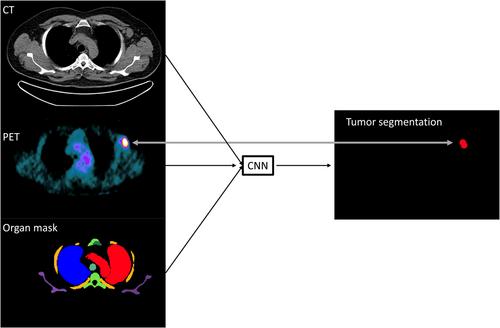
Forty-eight consecutive Hodgkin lymphoma (HL) patients staged with [18F] fluorodeoxyglucose positron emission tomography/computed tomography were included. The median age was 35 years (range: 7–75), 46% female. The tMTV was automatically measured using the AI-based tools positron emission tomography assisted reporting system (PARS) (from Siemens) and RECOMIA (recomia.org) without any manual adjustments. A group of eight nuclear medicine specialists manually segmented lesions for tMTV calculations; each patient was independently segmented by two specialists.
Results
The median of the manual tMTV was 146 cm3 (interquartile range [IQR]: 79–568 cm3) and the median difference between two tMTV values segmented by different specialists for the same patient was 26 cm3 (IQR: 10–86 cm3). In 22 of the 48 patients, the manual tMTV value was closer to the RECOMIA tMTV value than to the manual tMTV value segmented by the second specialist. In 11 of the remaining 26 patients, the difference between the RECOMIA tMTV and the manual tMTV was small (<26 cm3, which was the median difference between two manual tMTV values from the same patient). The corresponding numbers for PARS were 18 and 10 patients, respectively.
Conclusion
The results of this study indicate that RECOMIA and Siemens PARS AI tools could be used without any major manual adjustments in 69% (33/48) and 58% (28/48) of HL patients, respectively. This demonstrates the feasibility of using AI tools to support physicians measuring tMTV for assessment of prognosis in clinical practice.
期刊介绍:
Clinical Physiology and Functional Imaging publishes reports on clinical and experimental research pertinent to human physiology in health and disease. The scope of the Journal is very broad, covering all aspects of the regulatory system in the cardiovascular, renal and pulmonary systems with special emphasis on methodological aspects. The focus for the journal is, however, work that has potential clinical relevance. The Journal also features review articles on recent front-line research within these fields of interest.
Covered by the major abstracting services including Current Contents and Science Citation Index, Clinical Physiology and Functional Imaging plays an important role in providing effective and productive communication among clinical physiologists world-wide.
 求助内容:
求助内容: 应助结果提醒方式:


