{"title":"在行动中捕获:通过冷冻电子断层扫描细菌分泌系统的原位可视化。","authors":"Camille Keck, Jost Enninga, Léa Swistak","doi":"10.1111/mmi.15186","DOIUrl":null,"url":null,"abstract":"<p><p>Bacterial secretion systems, such as the type 3, 4, and 6 are multiprotein nanomachines expressed at the surface of pathogens with Gram-negative like envelopes. They are known to be crucial for virulence and to translocate bacteria-encoded effector proteins into host cells to manipulate cellular functions. This facilitates either pathogen attachment or invasion of the targeted cell. Effector proteins also promote evasion of host immune recognition. Imaging by cryo-electron microscopy in combination with structure determination has become a powerful approach to understand how these nanomachines work. Still, questions on their assembly, the precise secretion mechanisms, and their direct involvement in pathogenicity remain unsolved. Here, we present an overview of the recent developments in in situ cryo-electron microscopy. We discuss its potential for the investigation of the role of bacterial secretion systems during the host-bacterial crosstalk at the molecular level. These in situ studies open new perspectives for our understanding of secretion system structure and function.</p>","PeriodicalId":19006,"journal":{"name":"Molecular Microbiology","volume":null,"pages":null},"PeriodicalIF":2.6000,"publicationDate":"2024-04-01","publicationTypes":"Journal Article","fieldsOfStudy":null,"isOpenAccess":false,"openAccessPdf":"","citationCount":"0","resultStr":"{\"title\":\"Caught in the act: In situ visualization of bacterial secretion systems by cryo-electron tomography.\",\"authors\":\"Camille Keck, Jost Enninga, Léa Swistak\",\"doi\":\"10.1111/mmi.15186\",\"DOIUrl\":null,\"url\":null,\"abstract\":\"<p><p>Bacterial secretion systems, such as the type 3, 4, and 6 are multiprotein nanomachines expressed at the surface of pathogens with Gram-negative like envelopes. They are known to be crucial for virulence and to translocate bacteria-encoded effector proteins into host cells to manipulate cellular functions. This facilitates either pathogen attachment or invasion of the targeted cell. Effector proteins also promote evasion of host immune recognition. Imaging by cryo-electron microscopy in combination with structure determination has become a powerful approach to understand how these nanomachines work. Still, questions on their assembly, the precise secretion mechanisms, and their direct involvement in pathogenicity remain unsolved. Here, we present an overview of the recent developments in in situ cryo-electron microscopy. We discuss its potential for the investigation of the role of bacterial secretion systems during the host-bacterial crosstalk at the molecular level. These in situ studies open new perspectives for our understanding of secretion system structure and function.</p>\",\"PeriodicalId\":19006,\"journal\":{\"name\":\"Molecular Microbiology\",\"volume\":null,\"pages\":null},\"PeriodicalIF\":2.6000,\"publicationDate\":\"2024-04-01\",\"publicationTypes\":\"Journal Article\",\"fieldsOfStudy\":null,\"isOpenAccess\":false,\"openAccessPdf\":\"\",\"citationCount\":\"0\",\"resultStr\":null,\"platform\":\"Semanticscholar\",\"paperid\":null,\"PeriodicalName\":\"Molecular Microbiology\",\"FirstCategoryId\":\"99\",\"ListUrlMain\":\"https://doi.org/10.1111/mmi.15186\",\"RegionNum\":2,\"RegionCategory\":\"生物学\",\"ArticlePicture\":[],\"TitleCN\":null,\"AbstractTextCN\":null,\"PMCID\":null,\"EPubDate\":\"2023/11/17 0:00:00\",\"PubModel\":\"Epub\",\"JCR\":\"Q3\",\"JCRName\":\"BIOCHEMISTRY & MOLECULAR BIOLOGY\",\"Score\":null,\"Total\":0}","platform":"Semanticscholar","paperid":null,"PeriodicalName":"Molecular Microbiology","FirstCategoryId":"99","ListUrlMain":"https://doi.org/10.1111/mmi.15186","RegionNum":2,"RegionCategory":"生物学","ArticlePicture":[],"TitleCN":null,"AbstractTextCN":null,"PMCID":null,"EPubDate":"2023/11/17 0:00:00","PubModel":"Epub","JCR":"Q3","JCRName":"BIOCHEMISTRY & MOLECULAR BIOLOGY","Score":null,"Total":0}
Caught in the act: In situ visualization of bacterial secretion systems by cryo-electron tomography.
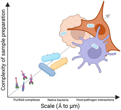
Bacterial secretion systems, such as the type 3, 4, and 6 are multiprotein nanomachines expressed at the surface of pathogens with Gram-negative like envelopes. They are known to be crucial for virulence and to translocate bacteria-encoded effector proteins into host cells to manipulate cellular functions. This facilitates either pathogen attachment or invasion of the targeted cell. Effector proteins also promote evasion of host immune recognition. Imaging by cryo-electron microscopy in combination with structure determination has become a powerful approach to understand how these nanomachines work. Still, questions on their assembly, the precise secretion mechanisms, and their direct involvement in pathogenicity remain unsolved. Here, we present an overview of the recent developments in in situ cryo-electron microscopy. We discuss its potential for the investigation of the role of bacterial secretion systems during the host-bacterial crosstalk at the molecular level. These in situ studies open new perspectives for our understanding of secretion system structure and function.
期刊介绍:
Molecular Microbiology, the leading primary journal in the microbial sciences, publishes molecular studies of Bacteria, Archaea, eukaryotic microorganisms, and their viruses.
Research papers should lead to a deeper understanding of the molecular principles underlying basic physiological processes or mechanisms. Appropriate topics include gene expression and regulation, pathogenicity and virulence, physiology and metabolism, synthesis of macromolecules (proteins, nucleic acids, lipids, polysaccharides, etc), cell biology and subcellular organization, membrane biogenesis and function, traffic and transport, cell-cell communication and signalling pathways, evolution and gene transfer. Articles focused on host responses (cellular or immunological) to pathogens or on microbial ecology should be directed to our sister journals Cellular Microbiology and Environmental Microbiology, respectively.
 求助内容:
求助内容: 应助结果提醒方式:


