{"title":"重建Pucapampella和鱼石螈的头下肌肉组织","authors":"Alexander N. Kuznetsov, Nadezhda V. Kryukova","doi":"10.1002/jmor.21648","DOIUrl":null,"url":null,"abstract":"<p>We present new reconstructions of subcephalic musculature for the stem chondrichthyan <i>Pucapampella</i>, the tetrapodomorph fish <i>Eusthenopteron</i>, and the Devonian tetrapod <i>Ichthyostega</i>. These reconstructions are based on macroscopic dissections of the head muscles of an archaic shark <i>Heptranchias</i> and an archaic actinopterygian <i>Polypterus</i>, that are combined with functional considerations and a reappraisal of not widely known theoretical concepts from the past. The subcephalic, as well as the supracephalic, musculature is formed by four anterior myomeres. They are continuous with subsequent myomeres of the trunk, but are innervated by ventral nerve roots of the <i>medulla oblongata</i> and thus belong to the head. The fourth subcephalic myomere ends with its posterior myoseptum on the occiput in osteichthyans, but on the first vertebra in chondrichthyans. The original function of subcephalic and supracephalic muscles in basal gnathostomes supposedly was to hold together anterior and posterior parts of the neurocranium during interaction with prey, such as the backward-ripping prey dissection, hypothesized for <i>Pucapampella</i>. In sarcopterygian osteichthyans, subcephalic musculature is involved in active depression of the anterior part of the neurocranium; specialization of this mechanism resulted in a complete separation of <i>m. subcephalicus</i> from trunk myomeres in <i>Latimeria</i>. Fusion of anterior and posterior parts of the neurocranium has resulted in reduction of the subcephalic musculature in the majority of cartilaginous and bony fishes. However, hexanchid sharks retain three posterior subcephalic myomeres for backward-ripping prey dissection. <i>Polypterus</i> and <i>Chauliodus</i> have retained the subcephalic musculature, but its function has shifted to a depression of the whole neurocranium.</p>","PeriodicalId":16528,"journal":{"name":"Journal of Morphology","volume":null,"pages":null},"PeriodicalIF":1.5000,"publicationDate":"2023-11-01","publicationTypes":"Journal Article","fieldsOfStudy":null,"isOpenAccess":false,"openAccessPdf":"","citationCount":"0","resultStr":"{\"title\":\"Reconstructing the subcephalic musculature in Pucapampella and Ichthyostega\",\"authors\":\"Alexander N. Kuznetsov, Nadezhda V. Kryukova\",\"doi\":\"10.1002/jmor.21648\",\"DOIUrl\":null,\"url\":null,\"abstract\":\"<p>We present new reconstructions of subcephalic musculature for the stem chondrichthyan <i>Pucapampella</i>, the tetrapodomorph fish <i>Eusthenopteron</i>, and the Devonian tetrapod <i>Ichthyostega</i>. These reconstructions are based on macroscopic dissections of the head muscles of an archaic shark <i>Heptranchias</i> and an archaic actinopterygian <i>Polypterus</i>, that are combined with functional considerations and a reappraisal of not widely known theoretical concepts from the past. The subcephalic, as well as the supracephalic, musculature is formed by four anterior myomeres. They are continuous with subsequent myomeres of the trunk, but are innervated by ventral nerve roots of the <i>medulla oblongata</i> and thus belong to the head. The fourth subcephalic myomere ends with its posterior myoseptum on the occiput in osteichthyans, but on the first vertebra in chondrichthyans. The original function of subcephalic and supracephalic muscles in basal gnathostomes supposedly was to hold together anterior and posterior parts of the neurocranium during interaction with prey, such as the backward-ripping prey dissection, hypothesized for <i>Pucapampella</i>. In sarcopterygian osteichthyans, subcephalic musculature is involved in active depression of the anterior part of the neurocranium; specialization of this mechanism resulted in a complete separation of <i>m. subcephalicus</i> from trunk myomeres in <i>Latimeria</i>. Fusion of anterior and posterior parts of the neurocranium has resulted in reduction of the subcephalic musculature in the majority of cartilaginous and bony fishes. However, hexanchid sharks retain three posterior subcephalic myomeres for backward-ripping prey dissection. <i>Polypterus</i> and <i>Chauliodus</i> have retained the subcephalic musculature, but its function has shifted to a depression of the whole neurocranium.</p>\",\"PeriodicalId\":16528,\"journal\":{\"name\":\"Journal of Morphology\",\"volume\":null,\"pages\":null},\"PeriodicalIF\":1.5000,\"publicationDate\":\"2023-11-01\",\"publicationTypes\":\"Journal Article\",\"fieldsOfStudy\":null,\"isOpenAccess\":false,\"openAccessPdf\":\"\",\"citationCount\":\"0\",\"resultStr\":null,\"platform\":\"Semanticscholar\",\"paperid\":null,\"PeriodicalName\":\"Journal of Morphology\",\"FirstCategoryId\":\"3\",\"ListUrlMain\":\"https://onlinelibrary.wiley.com/doi/10.1002/jmor.21648\",\"RegionNum\":4,\"RegionCategory\":\"医学\",\"ArticlePicture\":[],\"TitleCN\":null,\"AbstractTextCN\":null,\"PMCID\":null,\"EPubDate\":\"\",\"PubModel\":\"\",\"JCR\":\"Q2\",\"JCRName\":\"ANATOMY & MORPHOLOGY\",\"Score\":null,\"Total\":0}","platform":"Semanticscholar","paperid":null,"PeriodicalName":"Journal of Morphology","FirstCategoryId":"3","ListUrlMain":"https://onlinelibrary.wiley.com/doi/10.1002/jmor.21648","RegionNum":4,"RegionCategory":"医学","ArticlePicture":[],"TitleCN":null,"AbstractTextCN":null,"PMCID":null,"EPubDate":"","PubModel":"","JCR":"Q2","JCRName":"ANATOMY & MORPHOLOGY","Score":null,"Total":0}
Reconstructing the subcephalic musculature in Pucapampella and Ichthyostega
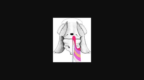
We present new reconstructions of subcephalic musculature for the stem chondrichthyan Pucapampella, the tetrapodomorph fish Eusthenopteron, and the Devonian tetrapod Ichthyostega. These reconstructions are based on macroscopic dissections of the head muscles of an archaic shark Heptranchias and an archaic actinopterygian Polypterus, that are combined with functional considerations and a reappraisal of not widely known theoretical concepts from the past. The subcephalic, as well as the supracephalic, musculature is formed by four anterior myomeres. They are continuous with subsequent myomeres of the trunk, but are innervated by ventral nerve roots of the medulla oblongata and thus belong to the head. The fourth subcephalic myomere ends with its posterior myoseptum on the occiput in osteichthyans, but on the first vertebra in chondrichthyans. The original function of subcephalic and supracephalic muscles in basal gnathostomes supposedly was to hold together anterior and posterior parts of the neurocranium during interaction with prey, such as the backward-ripping prey dissection, hypothesized for Pucapampella. In sarcopterygian osteichthyans, subcephalic musculature is involved in active depression of the anterior part of the neurocranium; specialization of this mechanism resulted in a complete separation of m. subcephalicus from trunk myomeres in Latimeria. Fusion of anterior and posterior parts of the neurocranium has resulted in reduction of the subcephalic musculature in the majority of cartilaginous and bony fishes. However, hexanchid sharks retain three posterior subcephalic myomeres for backward-ripping prey dissection. Polypterus and Chauliodus have retained the subcephalic musculature, but its function has shifted to a depression of the whole neurocranium.
期刊介绍:
The Journal of Morphology welcomes articles of original research in cytology, protozoology, embryology, and general morphology. Articles generally should not exceed 35 printed pages. Preliminary notices or articles of a purely descriptive morphological or taxonomic nature are not included. No paper which has already been published will be accepted, nor will simultaneous publications elsewhere be allowed.
The Journal of Morphology publishes research in functional, comparative, evolutionary and developmental morphology from vertebrates and invertebrates. Human and veterinary anatomy or paleontology are considered when an explicit connection to neontological animal morphology is presented, and the paper contains relevant information for the community of animal morphologists. Based on our long tradition, we continue to seek publishing the best papers in animal morphology.
 求助内容:
求助内容: 应助结果提醒方式:


