骨包虫病6例影像学表现及误诊分析。

IF 1.4 4区 医学 Q3 PARASITOLOGY
Yanqiu Sun, Chunlong Yan, Dengfeng Tian, Chenhong Zhang, Qiang Zhang
{"title":"骨包虫病6例影像学表现及误诊分析。","authors":"Yanqiu Sun, Chunlong Yan, Dengfeng Tian, Chenhong Zhang, Qiang Zhang","doi":"10.3347/kjp.2022.60.6.413","DOIUrl":null,"url":null,"abstract":"We retrospectively evaluated the clinical and imaging features of 6 patients with bone hydatid disease confirmed by surgery and pathological examination. Among the 6 patients, 2 were infected with Echinococcosis granulosus metacestode and 4 were infected with E. multilocularis metacestode. The 2 cases with cystic echinococcosis were diagnosed by computed tomographic (CT) examination, and other 4 cases were diagnosed by magnetic resonance (MR) imaging. On the initial evaluation, 1 case each was misdiagnosed as a giant cell tumor or neurogenic tumor, and 2 were misdiagnosed as tuberculosis. The imaging manifestations of bone hydatid disease are complex, but most common findings include expansive osteolytic bone destruction, which may be associated with sclerosing edges or dead bone formation, localized soft tissue masses, and vertebral lesions with wedge-shaped changes and spinal stenosis. Combining imaging findings with the patient’s epidemiological history and immunological examinations is of great help in improving the diagnosis and differential diagnosis of bone hydatid disease.","PeriodicalId":49938,"journal":{"name":"Korean Journal of Parasitology","volume":null,"pages":null},"PeriodicalIF":1.4000,"publicationDate":"2022-12-01","publicationTypes":"Journal Article","fieldsOfStudy":null,"isOpenAccess":false,"openAccessPdf":"https://ftp.ncbi.nlm.nih.gov/pub/pmc/oa_pdf/85/8d/kjp-60-6-413.PMC9806501.pdf","citationCount":"2","resultStr":"{\"title\":\"Imaging Manifestations and Misdiagnosis Analysis of Six Cases of Bone Hydatid Disease.\",\"authors\":\"Yanqiu Sun, Chunlong Yan, Dengfeng Tian, Chenhong Zhang, Qiang Zhang\",\"doi\":\"10.3347/kjp.2022.60.6.413\",\"DOIUrl\":null,\"url\":null,\"abstract\":\"We retrospectively evaluated the clinical and imaging features of 6 patients with bone hydatid disease confirmed by surgery and pathological examination. Among the 6 patients, 2 were infected with Echinococcosis granulosus metacestode and 4 were infected with E. multilocularis metacestode. The 2 cases with cystic echinococcosis were diagnosed by computed tomographic (CT) examination, and other 4 cases were diagnosed by magnetic resonance (MR) imaging. On the initial evaluation, 1 case each was misdiagnosed as a giant cell tumor or neurogenic tumor, and 2 were misdiagnosed as tuberculosis. The imaging manifestations of bone hydatid disease are complex, but most common findings include expansive osteolytic bone destruction, which may be associated with sclerosing edges or dead bone formation, localized soft tissue masses, and vertebral lesions with wedge-shaped changes and spinal stenosis. Combining imaging findings with the patient’s epidemiological history and immunological examinations is of great help in improving the diagnosis and differential diagnosis of bone hydatid disease.\",\"PeriodicalId\":49938,\"journal\":{\"name\":\"Korean Journal of Parasitology\",\"volume\":null,\"pages\":null},\"PeriodicalIF\":1.4000,\"publicationDate\":\"2022-12-01\",\"publicationTypes\":\"Journal Article\",\"fieldsOfStudy\":null,\"isOpenAccess\":false,\"openAccessPdf\":\"https://ftp.ncbi.nlm.nih.gov/pub/pmc/oa_pdf/85/8d/kjp-60-6-413.PMC9806501.pdf\",\"citationCount\":\"2\",\"resultStr\":null,\"platform\":\"Semanticscholar\",\"paperid\":null,\"PeriodicalName\":\"Korean Journal of Parasitology\",\"FirstCategoryId\":\"3\",\"ListUrlMain\":\"https://doi.org/10.3347/kjp.2022.60.6.413\",\"RegionNum\":4,\"RegionCategory\":\"医学\",\"ArticlePicture\":[],\"TitleCN\":null,\"AbstractTextCN\":null,\"PMCID\":null,\"EPubDate\":\"\",\"PubModel\":\"\",\"JCR\":\"Q3\",\"JCRName\":\"PARASITOLOGY\",\"Score\":null,\"Total\":0}","platform":"Semanticscholar","paperid":null,"PeriodicalName":"Korean Journal of Parasitology","FirstCategoryId":"3","ListUrlMain":"https://doi.org/10.3347/kjp.2022.60.6.413","RegionNum":4,"RegionCategory":"医学","ArticlePicture":[],"TitleCN":null,"AbstractTextCN":null,"PMCID":null,"EPubDate":"","PubModel":"","JCR":"Q3","JCRName":"PARASITOLOGY","Score":null,"Total":0}
引用次数: 2

摘要

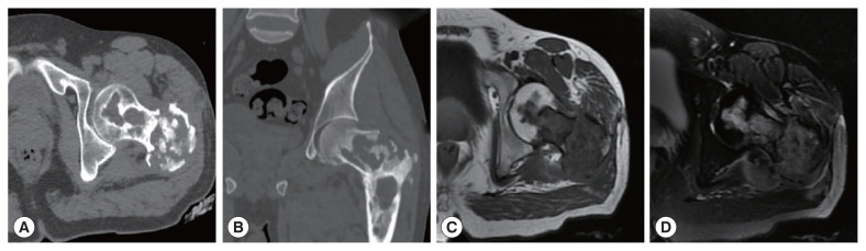
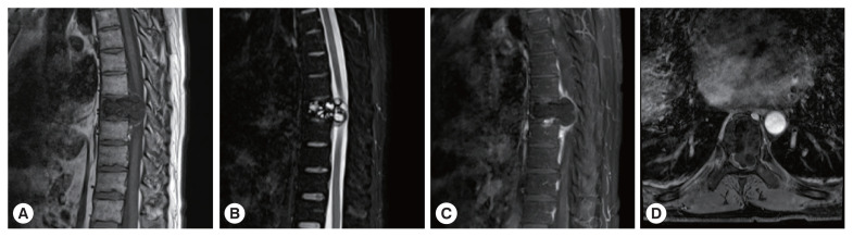
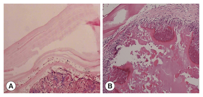
我们回顾性分析6例经手术及病理证实的骨包虫病的临床及影像学特点。6例患者中2例感染细粒棘球蚴,4例感染多房棘球蚴。囊性包虫病2例经计算机断层扫描(CT)诊断,另外4例经磁共振(MR)成像诊断。初诊误诊为巨细胞瘤和神经源性肿瘤各1例,误诊为结核2例。骨包虫病的影像学表现复杂,但最常见的表现包括扩散性溶骨破坏,可能伴有边缘硬化或死骨形成,局部软组织肿块,椎体病变伴楔形改变和椎管狭窄。将影像学表现与患者的流行病学史和免疫学检查相结合,对提高骨包虫病的诊断和鉴别诊断有很大帮助。
本文章由计算机程序翻译,如有差异,请以英文原文为准。

Imaging Manifestations and Misdiagnosis Analysis of Six Cases of Bone Hydatid Disease.

Imaging Manifestations and Misdiagnosis Analysis of Six Cases of Bone Hydatid Disease.

Imaging Manifestations and Misdiagnosis Analysis of Six Cases of Bone Hydatid Disease.

Imaging Manifestations and Misdiagnosis Analysis of Six Cases of Bone Hydatid Disease.
We retrospectively evaluated the clinical and imaging features of 6 patients with bone hydatid disease confirmed by surgery and pathological examination. Among the 6 patients, 2 were infected with Echinococcosis granulosus metacestode and 4 were infected with E. multilocularis metacestode. The 2 cases with cystic echinococcosis were diagnosed by computed tomographic (CT) examination, and other 4 cases were diagnosed by magnetic resonance (MR) imaging. On the initial evaluation, 1 case each was misdiagnosed as a giant cell tumor or neurogenic tumor, and 2 were misdiagnosed as tuberculosis. The imaging manifestations of bone hydatid disease are complex, but most common findings include expansive osteolytic bone destruction, which may be associated with sclerosing edges or dead bone formation, localized soft tissue masses, and vertebral lesions with wedge-shaped changes and spinal stenosis. Combining imaging findings with the patient’s epidemiological history and immunological examinations is of great help in improving the diagnosis and differential diagnosis of bone hydatid disease.
求助全文
通过发布文献求助,成功后即可免费获取论文全文。 去求助
来源期刊
CiteScore
2.80
自引率
0.00%
发文量
48
审稿时长
1 months
期刊介绍: The Korean Journal of Parasitology is the official journal paperless, on-line publication after Vol. 53, 2015 of The Korean Society for Parasitology and Tropical Medicine. Abbreviated title is ‘Korean J Parasitol’. It was launched in 1963. It contains original articles, case reports, brief communications, reviews or mini-reviews, book reviews, and letters to the editor on parasites of humans and animals, vectors, host-parasite relationships, zoonoses, and tropical medicine. It is published bimonthly in February, April, June, August, October, and December each year. Supplement numbers are at times published. All of the manuscripts are peer-reviewed.
×
引用
GB/T 7714-2015
复制
MLA
复制
APA
复制
导出至
BibTeX EndNote RefMan NoteFirst NoteExpress
×
提示
您的信息不完整,为了账户安全,请先补充。
现在去补充
×
提示
您因"违规操作"
具体请查看互助需知
我知道了
×
提示
确定
请完成安全验证×
copy
已复制链接
快去分享给好友吧!
我知道了
右上角分享
点击右上角分享
0
联系我们:info@booksci.cn Book学术提供免费学术资源搜索服务,方便国内外学者检索中英文文献。致力于提供最便捷和优质的服务体验。 Copyright © 2023 布克学术 All rights reserved.
京ICP备2023020795号-1
ghs 京公网安备 11010802042870号
Book学术文献互助
Book学术文献互助群
群 号:481959085
Book学术官方微信