Hülya Çetin Tunçez, Ali Murat Koç, Zehra Hilal Adıbelli, Fatma Zeynep Arslan, Asuman Argon, Gülşen Yücel Oğuzdoğan
{"title":"超声、多普勒超声及弹性成像对疑似恶性淋巴结的诊断价值。","authors":"Hülya Çetin Tunçez, Ali Murat Koç, Zehra Hilal Adıbelli, Fatma Zeynep Arslan, Asuman Argon, Gülşen Yücel Oğuzdoğan","doi":"10.15557/JoU.2023.0001","DOIUrl":null,"url":null,"abstract":"<p><strong>Aim: </strong>In this prospective study, the efficiency of imaging findings was investigated by comparing the histopathological results of lymph nodes with Doppler and ultrasound features and elasticity scores.</p><p><strong>Material and method: </strong>A total of 100 cervical or axillary lymph nodes with a suspected malignancy or whose size did not decrease after treatment were examined. In addition to the demographic data of the patients, B-mode ultrasound, Doppler ultrasound, and elastography features of the lymph nodes were evaluated prospectively. The irregular shape, increased size, pronounced hypoechogenicity, presence of micro/macro calcification, short axis/long axis ratio >2, increased size of the short axis, increased cortex thickness, obliterated hilus or increased cortex thickness >3.5 mm were evaluated on ultrasound. Resistivity index, pulsatility index, acceleration rate and time were evaluated for intranodal arterial structures on color. Doppler ultrasound, strain ratio value and elasticity score were recorded on ultrasound elastography. After sonographic examination, patients underwent ultrasound-guided fine needle aspiration cytology or tru-cutting needle biopsy. Histopathological examination results of the patients were compared with the B-mode ultrasound, Doppler ultrasound, and ultrasound elastography.</p><p><strong>Results: </strong>When the individual and combined effects of the ultrasound, Doppler ultrasound, and ultrasound elastography were evaluated, the combination of all three imaging methods was found to have the highest sensitivity and the highest overall accuracy (90.4% and 73.9%). As an individual method Doppler ultrasound had the highest specificity (77.8%). B-mode ultrasound was found to have the lowest accuracy (56.7%) both in individual and combined evaluations.</p><p><strong>Conclusion: </strong>Addition of ultrasound elastography to the combination of B-mode and Doppler ultrasound findings increases diagnostic sensitivity and accuracy in the differentiation of benign and malignant lymph nodes.</p>","PeriodicalId":45612,"journal":{"name":"Journal of Ultrasonography","volume":"23 92","pages":"1-9"},"PeriodicalIF":1.5000,"publicationDate":"2023-01-01","publicationTypes":"Journal Article","fieldsOfStudy":null,"isOpenAccess":false,"openAccessPdf":"https://ftp.ncbi.nlm.nih.gov/pub/pmc/oa_pdf/a3/3f/jou-23-001.PMC9985183.pdf","citationCount":"0","resultStr":"{\"title\":\"Diagnostic Efficacy of Ultrasonography, Doppler Ultrasonography and Elastography in the Evaluation of Suspected Malignant Lymph Nodes.\",\"authors\":\"Hülya Çetin Tunçez, Ali Murat Koç, Zehra Hilal Adıbelli, Fatma Zeynep Arslan, Asuman Argon, Gülşen Yücel Oğuzdoğan\",\"doi\":\"10.15557/JoU.2023.0001\",\"DOIUrl\":null,\"url\":null,\"abstract\":\"<p><strong>Aim: </strong>In this prospective study, the efficiency of imaging findings was investigated by comparing the histopathological results of lymph nodes with Doppler and ultrasound features and elasticity scores.</p><p><strong>Material and method: </strong>A total of 100 cervical or axillary lymph nodes with a suspected malignancy or whose size did not decrease after treatment were examined. In addition to the demographic data of the patients, B-mode ultrasound, Doppler ultrasound, and elastography features of the lymph nodes were evaluated prospectively. The irregular shape, increased size, pronounced hypoechogenicity, presence of micro/macro calcification, short axis/long axis ratio >2, increased size of the short axis, increased cortex thickness, obliterated hilus or increased cortex thickness >3.5 mm were evaluated on ultrasound. Resistivity index, pulsatility index, acceleration rate and time were evaluated for intranodal arterial structures on color. Doppler ultrasound, strain ratio value and elasticity score were recorded on ultrasound elastography. After sonographic examination, patients underwent ultrasound-guided fine needle aspiration cytology or tru-cutting needle biopsy. Histopathological examination results of the patients were compared with the B-mode ultrasound, Doppler ultrasound, and ultrasound elastography.</p><p><strong>Results: </strong>When the individual and combined effects of the ultrasound, Doppler ultrasound, and ultrasound elastography were evaluated, the combination of all three imaging methods was found to have the highest sensitivity and the highest overall accuracy (90.4% and 73.9%). As an individual method Doppler ultrasound had the highest specificity (77.8%). B-mode ultrasound was found to have the lowest accuracy (56.7%) both in individual and combined evaluations.</p><p><strong>Conclusion: </strong>Addition of ultrasound elastography to the combination of B-mode and Doppler ultrasound findings increases diagnostic sensitivity and accuracy in the differentiation of benign and malignant lymph nodes.</p>\",\"PeriodicalId\":45612,\"journal\":{\"name\":\"Journal of Ultrasonography\",\"volume\":\"23 92\",\"pages\":\"1-9\"},\"PeriodicalIF\":1.5000,\"publicationDate\":\"2023-01-01\",\"publicationTypes\":\"Journal Article\",\"fieldsOfStudy\":null,\"isOpenAccess\":false,\"openAccessPdf\":\"https://ftp.ncbi.nlm.nih.gov/pub/pmc/oa_pdf/a3/3f/jou-23-001.PMC9985183.pdf\",\"citationCount\":\"0\",\"resultStr\":null,\"platform\":\"Semanticscholar\",\"paperid\":null,\"PeriodicalName\":\"Journal of Ultrasonography\",\"FirstCategoryId\":\"1085\",\"ListUrlMain\":\"https://doi.org/10.15557/JoU.2023.0001\",\"RegionNum\":0,\"RegionCategory\":null,\"ArticlePicture\":[],\"TitleCN\":null,\"AbstractTextCN\":null,\"PMCID\":null,\"EPubDate\":\"\",\"PubModel\":\"\",\"JCR\":\"Q3\",\"JCRName\":\"RADIOLOGY, NUCLEAR MEDICINE & MEDICAL IMAGING\",\"Score\":null,\"Total\":0}","platform":"Semanticscholar","paperid":null,"PeriodicalName":"Journal of Ultrasonography","FirstCategoryId":"1085","ListUrlMain":"https://doi.org/10.15557/JoU.2023.0001","RegionNum":0,"RegionCategory":null,"ArticlePicture":[],"TitleCN":null,"AbstractTextCN":null,"PMCID":null,"EPubDate":"","PubModel":"","JCR":"Q3","JCRName":"RADIOLOGY, NUCLEAR MEDICINE & MEDICAL IMAGING","Score":null,"Total":0}
Diagnostic Efficacy of Ultrasonography, Doppler Ultrasonography and Elastography in the Evaluation of Suspected Malignant Lymph Nodes.
Aim: In this prospective study, the efficiency of imaging findings was investigated by comparing the histopathological results of lymph nodes with Doppler and ultrasound features and elasticity scores.
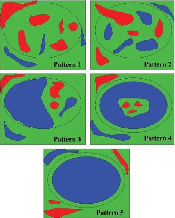
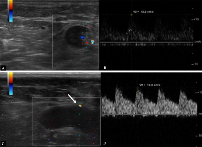
Material and method: A total of 100 cervical or axillary lymph nodes with a suspected malignancy or whose size did not decrease after treatment were examined. In addition to the demographic data of the patients, B-mode ultrasound, Doppler ultrasound, and elastography features of the lymph nodes were evaluated prospectively. The irregular shape, increased size, pronounced hypoechogenicity, presence of micro/macro calcification, short axis/long axis ratio >2, increased size of the short axis, increased cortex thickness, obliterated hilus or increased cortex thickness >3.5 mm were evaluated on ultrasound. Resistivity index, pulsatility index, acceleration rate and time were evaluated for intranodal arterial structures on color. Doppler ultrasound, strain ratio value and elasticity score were recorded on ultrasound elastography. After sonographic examination, patients underwent ultrasound-guided fine needle aspiration cytology or tru-cutting needle biopsy. Histopathological examination results of the patients were compared with the B-mode ultrasound, Doppler ultrasound, and ultrasound elastography.
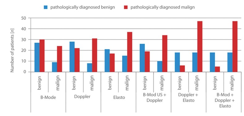
Results: When the individual and combined effects of the ultrasound, Doppler ultrasound, and ultrasound elastography were evaluated, the combination of all three imaging methods was found to have the highest sensitivity and the highest overall accuracy (90.4% and 73.9%). As an individual method Doppler ultrasound had the highest specificity (77.8%). B-mode ultrasound was found to have the lowest accuracy (56.7%) both in individual and combined evaluations.
Conclusion: Addition of ultrasound elastography to the combination of B-mode and Doppler ultrasound findings increases diagnostic sensitivity and accuracy in the differentiation of benign and malignant lymph nodes.

 求助内容:
求助内容: 应助结果提醒方式:


