Yan-Yun Cao, Jing Ning, Ru-Zhi Zhang, Kang Ge, Ting-Ting Huang
{"title":"CM-Dil 标记的 Muse 细胞在大鼠培养和皮肤伤口中的特性。","authors":"Yan-Yun Cao, Jing Ning, Ru-Zhi Zhang, Kang Ge, Ting-Ting Huang","doi":"10.1007/s10561-022-10067-9","DOIUrl":null,"url":null,"abstract":"<p><p>To investigate the characteristics of multilineage-differentiating stress-enduring (Muse) cells labeled with chloromethyl dialkylcarbocyanine (CM-Dil) in culture and in skin wounds of rats. Normal human dermal fibroblasts (NHDFs) were obtained from foreskins and were confirmed by immunocytochemistry with vimentin. Muse cells were derived from NHDFs using long-term trypsinization (LTT), were confirmed using immunocytochemistry with antibodies against stage specific embryonic antigen-3 (SSEA-3) and CD105 and were expanded in suspension cultures. The Muse cells were labeled with CM-Dil and were further evaluated with respect to their biological properties using CCK-8 assays and scratch tests. One hundred µl CM-Dil-labeled Muse cells at a concentration of 5 × 10<sup>3</sup>/µl were injected subcutaneously at the edges of skin wounds in adult male SD rats. At weeks 1, 3 and 5 after the injection, the distribution of CM-Dil-labeled Muse cells in skin tissues was observed using immunofluorescence microscopy. Muse cells were double-positive for CD105 and SSEA-3. ALP staining of the M-clusters were positive and they displayed orange-red fluorescence after labelling with CM-Dil, which had no adverse effects on their viability, migration or differentiation capacity. One week after the subcutaneous injection of CM-Dil-labeled Muse cells, many cells with orange-red fluorescence were observed at the edges of the skin injuries; those fluorescent spots gradually decreased over time, and only a few Muse cells with fluorescence could be detected by week 5. CM-Dil can be used to label Muse cells without affecting their proliferation, migration or differentiation, and can be used for short-term tracking of Muse cells for the treatment of skin wounds in a rat model.</p>","PeriodicalId":9723,"journal":{"name":"Cell and Tissue Banking","volume":" ","pages":"285-294"},"PeriodicalIF":1.4000,"publicationDate":"2024-03-01","publicationTypes":"Journal Article","fieldsOfStudy":null,"isOpenAccess":false,"openAccessPdf":"","citationCount":"0","resultStr":"{\"title\":\"Characterization of CM-Dil-labeled Muse cells in culture and in skin wounds in rats.\",\"authors\":\"Yan-Yun Cao, Jing Ning, Ru-Zhi Zhang, Kang Ge, Ting-Ting Huang\",\"doi\":\"10.1007/s10561-022-10067-9\",\"DOIUrl\":null,\"url\":null,\"abstract\":\"<p><p>To investigate the characteristics of multilineage-differentiating stress-enduring (Muse) cells labeled with chloromethyl dialkylcarbocyanine (CM-Dil) in culture and in skin wounds of rats. Normal human dermal fibroblasts (NHDFs) were obtained from foreskins and were confirmed by immunocytochemistry with vimentin. Muse cells were derived from NHDFs using long-term trypsinization (LTT), were confirmed using immunocytochemistry with antibodies against stage specific embryonic antigen-3 (SSEA-3) and CD105 and were expanded in suspension cultures. The Muse cells were labeled with CM-Dil and were further evaluated with respect to their biological properties using CCK-8 assays and scratch tests. One hundred µl CM-Dil-labeled Muse cells at a concentration of 5 × 10<sup>3</sup>/µl were injected subcutaneously at the edges of skin wounds in adult male SD rats. At weeks 1, 3 and 5 after the injection, the distribution of CM-Dil-labeled Muse cells in skin tissues was observed using immunofluorescence microscopy. Muse cells were double-positive for CD105 and SSEA-3. ALP staining of the M-clusters were positive and they displayed orange-red fluorescence after labelling with CM-Dil, which had no adverse effects on their viability, migration or differentiation capacity. One week after the subcutaneous injection of CM-Dil-labeled Muse cells, many cells with orange-red fluorescence were observed at the edges of the skin injuries; those fluorescent spots gradually decreased over time, and only a few Muse cells with fluorescence could be detected by week 5. CM-Dil can be used to label Muse cells without affecting their proliferation, migration or differentiation, and can be used for short-term tracking of Muse cells for the treatment of skin wounds in a rat model.</p>\",\"PeriodicalId\":9723,\"journal\":{\"name\":\"Cell and Tissue Banking\",\"volume\":\" \",\"pages\":\"285-294\"},\"PeriodicalIF\":1.4000,\"publicationDate\":\"2024-03-01\",\"publicationTypes\":\"Journal Article\",\"fieldsOfStudy\":null,\"isOpenAccess\":false,\"openAccessPdf\":\"\",\"citationCount\":\"0\",\"resultStr\":null,\"platform\":\"Semanticscholar\",\"paperid\":null,\"PeriodicalName\":\"Cell and Tissue Banking\",\"FirstCategoryId\":\"5\",\"ListUrlMain\":\"https://doi.org/10.1007/s10561-022-10067-9\",\"RegionNum\":4,\"RegionCategory\":\"医学\",\"ArticlePicture\":[],\"TitleCN\":null,\"AbstractTextCN\":null,\"PMCID\":null,\"EPubDate\":\"2023/1/8 0:00:00\",\"PubModel\":\"Epub\",\"JCR\":\"Q4\",\"JCRName\":\"CELL BIOLOGY\",\"Score\":null,\"Total\":0}","platform":"Semanticscholar","paperid":null,"PeriodicalName":"Cell and Tissue Banking","FirstCategoryId":"5","ListUrlMain":"https://doi.org/10.1007/s10561-022-10067-9","RegionNum":4,"RegionCategory":"医学","ArticlePicture":[],"TitleCN":null,"AbstractTextCN":null,"PMCID":null,"EPubDate":"2023/1/8 0:00:00","PubModel":"Epub","JCR":"Q4","JCRName":"CELL BIOLOGY","Score":null,"Total":0}
Characterization of CM-Dil-labeled Muse cells in culture and in skin wounds in rats.
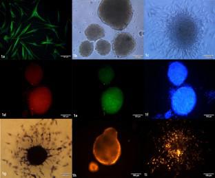
To investigate the characteristics of multilineage-differentiating stress-enduring (Muse) cells labeled with chloromethyl dialkylcarbocyanine (CM-Dil) in culture and in skin wounds of rats. Normal human dermal fibroblasts (NHDFs) were obtained from foreskins and were confirmed by immunocytochemistry with vimentin. Muse cells were derived from NHDFs using long-term trypsinization (LTT), were confirmed using immunocytochemistry with antibodies against stage specific embryonic antigen-3 (SSEA-3) and CD105 and were expanded in suspension cultures. The Muse cells were labeled with CM-Dil and were further evaluated with respect to their biological properties using CCK-8 assays and scratch tests. One hundred µl CM-Dil-labeled Muse cells at a concentration of 5 × 10³/µl were injected subcutaneously at the edges of skin wounds in adult male SD rats. At weeks 1, 3 and 5 after the injection, the distribution of CM-Dil-labeled Muse cells in skin tissues was observed using immunofluorescence microscopy. Muse cells were double-positive for CD105 and SSEA-3. ALP staining of the M-clusters were positive and they displayed orange-red fluorescence after labelling with CM-Dil, which had no adverse effects on their viability, migration or differentiation capacity. One week after the subcutaneous injection of CM-Dil-labeled Muse cells, many cells with orange-red fluorescence were observed at the edges of the skin injuries; those fluorescent spots gradually decreased over time, and only a few Muse cells with fluorescence could be detected by week 5. CM-Dil can be used to label Muse cells without affecting their proliferation, migration or differentiation, and can be used for short-term tracking of Muse cells for the treatment of skin wounds in a rat model.
期刊介绍:
Cell and Tissue Banking provides a forum for disseminating information to scientists and clinicians involved in the banking and transplantation of cells and tissues. Cell and Tissue Banking is an international, peer-reviewed journal that publishes original papers in the following areas:
basic research concerning general aspects of tissue banking such as quality assurance and control of banked cells/tissues, effects of preservation and sterilisation methods on cells/tissues, biotechnology, etc.; clinical applications of banked cells/tissues; standards of practice in procurement, processing, storage and distribution of cells/tissues; ethical issues; medico-legal issues.
 求助内容:
求助内容: 应助结果提醒方式:


