{"title":"全脑单纤维示踪揭示桥脚核靶皮质锥体神经元皮层下侧枝优先投射。","authors":"Qiao-Qiong Liu, Yu-Xiao Cheng, Qi Jing, Ke-Ming Zhang, Lu-Feng Ding, Xiao-Wei Fan, Chun-Hui Jia, Fang Xu, Guo-Qiang Bi, Pak-Ming Lau","doi":"10.1186/s13041-022-00975-y","DOIUrl":null,"url":null,"abstract":"<p><p>The pedunculopontine nucleus (PPN) is a heterogeneous midbrain structure involved in various brain functions, such as motor control, learning, reward, and sleep. Previous studies using conventional tracers have shown that the PPN receives extensive afferent inputs from various cortical areas. To examine how these cortical axons make collateral projections to other subcortical areas, we used a dual-viral injection strategy to sparsely label PPN-targeting cortical pyramidal neurons in CaMKIIα-Cre transgenic mice. Using a high-speed volumetric imaging with on-the-fly-scan and Readout (VISoR) technique, we visualized brain-wide axonal projections of individual PPN-targeting neurons from several cortical areas, including the prelimbic region (PL), anterior cingulate area (ACA) and secondary motor cortex (MOs). We found that each PPN-projecting neuron had a unique profile of collateralization, with some subcortical areas being preferential targets. In particular, PPN-projecting neurons from all three traced cortical areas exhibited common preferential collateralization to several nuclei, with most neurons targeting the striatum (STR), lateral hypothalamic area (LHA) and periaqueductal gray (PAG), and a substantial portion of neurons also targeting the zona incerta (ZI), median raphe nucleus (MRN) and substantia nigra pars reticulata (SNr). Meanwhile, very specific collateralization patterns were found for other nuclei, including the intermediate reticular nucleus (IRN), parvicellular reticular nucleus (PARN) and gigantocellular reticular nucleus (GRN), which receive collateral inputs almost exclusively from the MOs. These observations provide potential anatomical mechanisms for cortical neurons to coordinate the PPN with other subcortical areas in performing different physiological functions.</p>","PeriodicalId":18851,"journal":{"name":"Molecular Brain","volume":"15 1","pages":"88"},"PeriodicalIF":2.9000,"publicationDate":"2022-10-29","publicationTypes":"Journal Article","fieldsOfStudy":null,"isOpenAccess":false,"openAccessPdf":"https://www.ncbi.nlm.nih.gov/pmc/articles/PMC9618196/pdf/","citationCount":"2","resultStr":"{\"title\":\"Preferential subcortical collateral projections of pedunculopontine nucleus-targeting cortical pyramidal neurons revealed by brain-wide single fiber tracing.\",\"authors\":\"Qiao-Qiong Liu, Yu-Xiao Cheng, Qi Jing, Ke-Ming Zhang, Lu-Feng Ding, Xiao-Wei Fan, Chun-Hui Jia, Fang Xu, Guo-Qiang Bi, Pak-Ming Lau\",\"doi\":\"10.1186/s13041-022-00975-y\",\"DOIUrl\":null,\"url\":null,\"abstract\":\"<p><p>The pedunculopontine nucleus (PPN) is a heterogeneous midbrain structure involved in various brain functions, such as motor control, learning, reward, and sleep. Previous studies using conventional tracers have shown that the PPN receives extensive afferent inputs from various cortical areas. To examine how these cortical axons make collateral projections to other subcortical areas, we used a dual-viral injection strategy to sparsely label PPN-targeting cortical pyramidal neurons in CaMKIIα-Cre transgenic mice. Using a high-speed volumetric imaging with on-the-fly-scan and Readout (VISoR) technique, we visualized brain-wide axonal projections of individual PPN-targeting neurons from several cortical areas, including the prelimbic region (PL), anterior cingulate area (ACA) and secondary motor cortex (MOs). We found that each PPN-projecting neuron had a unique profile of collateralization, with some subcortical areas being preferential targets. In particular, PPN-projecting neurons from all three traced cortical areas exhibited common preferential collateralization to several nuclei, with most neurons targeting the striatum (STR), lateral hypothalamic area (LHA) and periaqueductal gray (PAG), and a substantial portion of neurons also targeting the zona incerta (ZI), median raphe nucleus (MRN) and substantia nigra pars reticulata (SNr). Meanwhile, very specific collateralization patterns were found for other nuclei, including the intermediate reticular nucleus (IRN), parvicellular reticular nucleus (PARN) and gigantocellular reticular nucleus (GRN), which receive collateral inputs almost exclusively from the MOs. These observations provide potential anatomical mechanisms for cortical neurons to coordinate the PPN with other subcortical areas in performing different physiological functions.</p>\",\"PeriodicalId\":18851,\"journal\":{\"name\":\"Molecular Brain\",\"volume\":\"15 1\",\"pages\":\"88\"},\"PeriodicalIF\":2.9000,\"publicationDate\":\"2022-10-29\",\"publicationTypes\":\"Journal Article\",\"fieldsOfStudy\":null,\"isOpenAccess\":false,\"openAccessPdf\":\"https://www.ncbi.nlm.nih.gov/pmc/articles/PMC9618196/pdf/\",\"citationCount\":\"2\",\"resultStr\":null,\"platform\":\"Semanticscholar\",\"paperid\":null,\"PeriodicalName\":\"Molecular Brain\",\"FirstCategoryId\":\"3\",\"ListUrlMain\":\"https://doi.org/10.1186/s13041-022-00975-y\",\"RegionNum\":3,\"RegionCategory\":\"医学\",\"ArticlePicture\":[],\"TitleCN\":null,\"AbstractTextCN\":null,\"PMCID\":null,\"EPubDate\":\"\",\"PubModel\":\"\",\"JCR\":\"Q2\",\"JCRName\":\"NEUROSCIENCES\",\"Score\":null,\"Total\":0}","platform":"Semanticscholar","paperid":null,"PeriodicalName":"Molecular Brain","FirstCategoryId":"3","ListUrlMain":"https://doi.org/10.1186/s13041-022-00975-y","RegionNum":3,"RegionCategory":"医学","ArticlePicture":[],"TitleCN":null,"AbstractTextCN":null,"PMCID":null,"EPubDate":"","PubModel":"","JCR":"Q2","JCRName":"NEUROSCIENCES","Score":null,"Total":0}
Preferential subcortical collateral projections of pedunculopontine nucleus-targeting cortical pyramidal neurons revealed by brain-wide single fiber tracing.
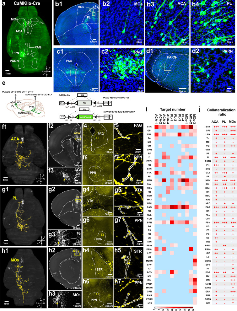
The pedunculopontine nucleus (PPN) is a heterogeneous midbrain structure involved in various brain functions, such as motor control, learning, reward, and sleep. Previous studies using conventional tracers have shown that the PPN receives extensive afferent inputs from various cortical areas. To examine how these cortical axons make collateral projections to other subcortical areas, we used a dual-viral injection strategy to sparsely label PPN-targeting cortical pyramidal neurons in CaMKIIα-Cre transgenic mice. Using a high-speed volumetric imaging with on-the-fly-scan and Readout (VISoR) technique, we visualized brain-wide axonal projections of individual PPN-targeting neurons from several cortical areas, including the prelimbic region (PL), anterior cingulate area (ACA) and secondary motor cortex (MOs). We found that each PPN-projecting neuron had a unique profile of collateralization, with some subcortical areas being preferential targets. In particular, PPN-projecting neurons from all three traced cortical areas exhibited common preferential collateralization to several nuclei, with most neurons targeting the striatum (STR), lateral hypothalamic area (LHA) and periaqueductal gray (PAG), and a substantial portion of neurons also targeting the zona incerta (ZI), median raphe nucleus (MRN) and substantia nigra pars reticulata (SNr). Meanwhile, very specific collateralization patterns were found for other nuclei, including the intermediate reticular nucleus (IRN), parvicellular reticular nucleus (PARN) and gigantocellular reticular nucleus (GRN), which receive collateral inputs almost exclusively from the MOs. These observations provide potential anatomical mechanisms for cortical neurons to coordinate the PPN with other subcortical areas in performing different physiological functions.
期刊介绍:
Molecular Brain is an open access, peer-reviewed journal that considers manuscripts on all aspects of studies on the nervous system at the molecular, cellular, and systems level providing a forum for scientists to communicate their findings.
Molecular brain research is a rapidly expanding research field in which integrative approaches at the genetic, molecular, cellular and synaptic levels yield key information about the physiological and pathological brain. These studies involve the use of a wide range of modern techniques in molecular biology, genomics, proteomics, imaging and electrophysiology.
 求助内容:
求助内容: 应助结果提醒方式:


