{"title":"利用三维定量参数图同时进行动脉和静脉成像。","authors":"Tomoki Amemiya, Suguru Yokosawa, Yo Taniguchi, Ryota Sato, Yoshihisa Soutome, Hisaaki Ochi, Toru Shirai","doi":"10.2463/mrms.mp.2021-0170","DOIUrl":null,"url":null,"abstract":"<p><strong>Purpose: </strong>To increase the number of images that can be acquired in MR examinations using quantitative parameters, we developed a method for obtaining arterial and venous images with mapping of proton density (PD), RF inhomogeneity (B1), longitudinal relaxation time (T1), apparent transverse relaxation time (T2*), and magnetic susceptibility through calculation, all with the same spatial resolution.</p><p><strong>Methods: </strong>The proposed method uses partially RF-spoiled gradient echo sequences to obtain 3D images of a subject with multiple scan parameters. The PD, B1, T1, T2*, and magnetic susceptibility maps are estimated using the quantification method we previously developed. Arterial images are obtained by adding images using optimized weights to emphasize the arteries. A morphology filter is used to obtain venous images from the magnetic susceptibility maps. For evaluation, images obtained from four out of five healthy volunteers were used to optimize the weights used in the arterial-image calculation, and the optimized weights were applied to the images from the fifth volunteer to obtain an arterial image. Arterial images of the five volunteers were calculated using the leave-one-out method, and the contrast between the arterial and background regions defined using the reference time-of-flight (TOF) method was evaluated using the area under the receiver operation characteristic curve (AUC). The contrast between venous and background regions defined by a reference quantitative susceptibility mapping (QSM) method was also evaluated for the venous image.</p><p><strong>Results: </strong>The AUC to discriminate blood vessels and background using the proposed method was 0.905 for the arterial image and 0.920 for the venous image.</p><p><strong>Conclusion: </strong>The results indicate that the arterial images and venous images have high signal intensity at the same region as determined from the reference TOF and QSM methods, demonstrating the possibility of acquiring vasculature images with quantitative parameter mapping through calculation in an integrated manner.</p>","PeriodicalId":18119,"journal":{"name":"Magnetic Resonance in Medical Sciences","volume":" ","pages":"56-65"},"PeriodicalIF":3.2000,"publicationDate":"2024-01-01","publicationTypes":"Journal Article","fieldsOfStudy":null,"isOpenAccess":false,"openAccessPdf":"https://www.ncbi.nlm.nih.gov/pmc/articles/PMC10838721/pdf/","citationCount":"0","resultStr":"{\"title\":\"Simultaneous Arterial and Venous Imaging Using 3D Quantitative Parameter Mapping.\",\"authors\":\"Tomoki Amemiya, Suguru Yokosawa, Yo Taniguchi, Ryota Sato, Yoshihisa Soutome, Hisaaki Ochi, Toru Shirai\",\"doi\":\"10.2463/mrms.mp.2021-0170\",\"DOIUrl\":null,\"url\":null,\"abstract\":\"<p><strong>Purpose: </strong>To increase the number of images that can be acquired in MR examinations using quantitative parameters, we developed a method for obtaining arterial and venous images with mapping of proton density (PD), RF inhomogeneity (B1), longitudinal relaxation time (T1), apparent transverse relaxation time (T2*), and magnetic susceptibility through calculation, all with the same spatial resolution.</p><p><strong>Methods: </strong>The proposed method uses partially RF-spoiled gradient echo sequences to obtain 3D images of a subject with multiple scan parameters. The PD, B1, T1, T2*, and magnetic susceptibility maps are estimated using the quantification method we previously developed. Arterial images are obtained by adding images using optimized weights to emphasize the arteries. A morphology filter is used to obtain venous images from the magnetic susceptibility maps. For evaluation, images obtained from four out of five healthy volunteers were used to optimize the weights used in the arterial-image calculation, and the optimized weights were applied to the images from the fifth volunteer to obtain an arterial image. Arterial images of the five volunteers were calculated using the leave-one-out method, and the contrast between the arterial and background regions defined using the reference time-of-flight (TOF) method was evaluated using the area under the receiver operation characteristic curve (AUC). The contrast between venous and background regions defined by a reference quantitative susceptibility mapping (QSM) method was also evaluated for the venous image.</p><p><strong>Results: </strong>The AUC to discriminate blood vessels and background using the proposed method was 0.905 for the arterial image and 0.920 for the venous image.</p><p><strong>Conclusion: </strong>The results indicate that the arterial images and venous images have high signal intensity at the same region as determined from the reference TOF and QSM methods, demonstrating the possibility of acquiring vasculature images with quantitative parameter mapping through calculation in an integrated manner.</p>\",\"PeriodicalId\":18119,\"journal\":{\"name\":\"Magnetic Resonance in Medical Sciences\",\"volume\":\" \",\"pages\":\"56-65\"},\"PeriodicalIF\":3.2000,\"publicationDate\":\"2024-01-01\",\"publicationTypes\":\"Journal Article\",\"fieldsOfStudy\":null,\"isOpenAccess\":false,\"openAccessPdf\":\"https://www.ncbi.nlm.nih.gov/pmc/articles/PMC10838721/pdf/\",\"citationCount\":\"0\",\"resultStr\":null,\"platform\":\"Semanticscholar\",\"paperid\":null,\"PeriodicalName\":\"Magnetic Resonance in Medical Sciences\",\"FirstCategoryId\":\"3\",\"ListUrlMain\":\"https://doi.org/10.2463/mrms.mp.2021-0170\",\"RegionNum\":3,\"RegionCategory\":\"医学\",\"ArticlePicture\":[],\"TitleCN\":null,\"AbstractTextCN\":null,\"PMCID\":null,\"EPubDate\":\"2022/12/21 0:00:00\",\"PubModel\":\"Epub\",\"JCR\":\"Q2\",\"JCRName\":\"RADIOLOGY, NUCLEAR MEDICINE & MEDICAL IMAGING\",\"Score\":null,\"Total\":0}","platform":"Semanticscholar","paperid":null,"PeriodicalName":"Magnetic Resonance in Medical Sciences","FirstCategoryId":"3","ListUrlMain":"https://doi.org/10.2463/mrms.mp.2021-0170","RegionNum":3,"RegionCategory":"医学","ArticlePicture":[],"TitleCN":null,"AbstractTextCN":null,"PMCID":null,"EPubDate":"2022/12/21 0:00:00","PubModel":"Epub","JCR":"Q2","JCRName":"RADIOLOGY, NUCLEAR MEDICINE & MEDICAL IMAGING","Score":null,"Total":0}
Simultaneous Arterial and Venous Imaging Using 3D Quantitative Parameter Mapping.
Purpose: To increase the number of images that can be acquired in MR examinations using quantitative parameters, we developed a method for obtaining arterial and venous images with mapping of proton density (PD), RF inhomogeneity (B1), longitudinal relaxation time (T1), apparent transverse relaxation time (T2*), and magnetic susceptibility through calculation, all with the same spatial resolution.
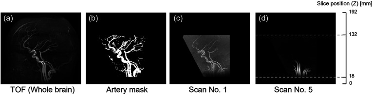
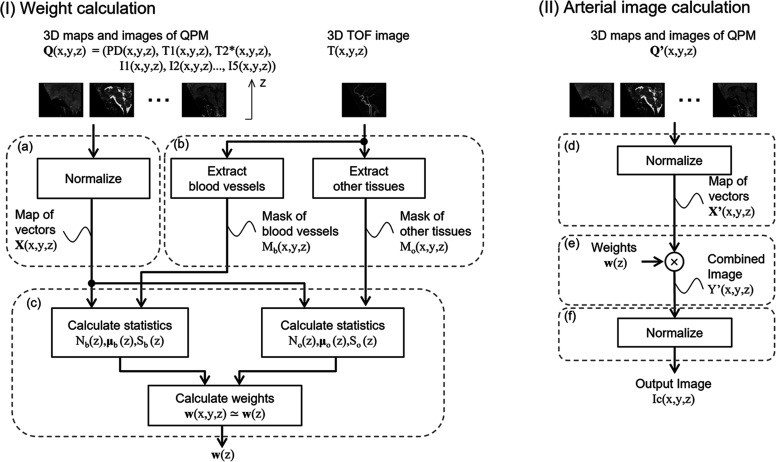
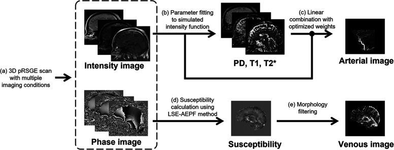
Methods: The proposed method uses partially RF-spoiled gradient echo sequences to obtain 3D images of a subject with multiple scan parameters. The PD, B1, T1, T2*, and magnetic susceptibility maps are estimated using the quantification method we previously developed. Arterial images are obtained by adding images using optimized weights to emphasize the arteries. A morphology filter is used to obtain venous images from the magnetic susceptibility maps. For evaluation, images obtained from four out of five healthy volunteers were used to optimize the weights used in the arterial-image calculation, and the optimized weights were applied to the images from the fifth volunteer to obtain an arterial image. Arterial images of the five volunteers were calculated using the leave-one-out method, and the contrast between the arterial and background regions defined using the reference time-of-flight (TOF) method was evaluated using the area under the receiver operation characteristic curve (AUC). The contrast between venous and background regions defined by a reference quantitative susceptibility mapping (QSM) method was also evaluated for the venous image.
Results: The AUC to discriminate blood vessels and background using the proposed method was 0.905 for the arterial image and 0.920 for the venous image.
Conclusion: The results indicate that the arterial images and venous images have high signal intensity at the same region as determined from the reference TOF and QSM methods, demonstrating the possibility of acquiring vasculature images with quantitative parameter mapping through calculation in an integrated manner.
期刊介绍:
Magnetic Resonance in Medical Sciences (MRMS or Magn
Reson Med Sci) is an international journal pursuing the
publication of original articles contributing to the progress
of magnetic resonance in the field of biomedical sciences
including technical developments and clinical applications.
MRMS is an official journal of the Japanese Society for
Magnetic Resonance in Medicine (JSMRM).
 求助内容:
求助内容: 应助结果提醒方式:


