Young Hee Jung, Seongbeom Park, Na Kyung Lee, Hyun Jeong Han, Hyemin Jang, Hee Jin Kim, Sang Won Seo, Duk Lyul Na
{"title":"白质病变主要位于深部白质代表栓塞病因,而不是小血管疾病。","authors":"Young Hee Jung, Seongbeom Park, Na Kyung Lee, Hyun Jeong Han, Hyemin Jang, Hee Jin Kim, Sang Won Seo, Duk Lyul Na","doi":"10.12779/dnd.2023.22.1.28","DOIUrl":null,"url":null,"abstract":"<p><strong>Background and purpose: </strong>We investigated the correlation between the deep distribution of white matter hyperintensity (WMH) (dWMH: WMH in deep and corticomedullary areas, with minimal periventricular WMH) and a positive agitated saline contrast echocardiography result.</p><p><strong>Methods: </strong>We retrospectively recruited participants with comprehensive dementia evaluations, an agitated saline study, and brain imaging. The participants were classified into two groups according to WMH-distributions: dWMH and dpWMH (mainly periventricular WMH with or without deep WMH.) We hypothesized that dWMH is more likely associated with embolism, whereas dpWMH is associated with small-vessel diseases. We compared the clinical characteristics, WMH-distributions, and positive rate of agitated saline studies between the two groups.</p><p><strong>Results: </strong>Among 90 participants, 27 and 12 met the dWMH and dpWMH criteria, respectively. The dWMH-group was younger (62.2±7.5 vs. 78.9±7.3, <i>p</i><0.001) and had a lower prevalence of hypertension (29.6% vs. 75%, <i>p</i>=0.008), diabetes mellitus (3.7% vs. 25%, <i>p</i>=0.043), and hyperlipidemia (33.3% vs. 83.3%, <i>p</i>=0.043) than the dpWMH-group. Regarding deep white matter lesions, the number of small lesions (<3 mm) was higher in the dWMH-group(10.9±9.7) than in the dpWMH-group (3.1±6.4) (<i>p</i>=0.008), and WMH was predominantly distributed in the border-zones and corticomedullary areas. Most importantly, the positive agitated saline study rate was higher in the dWMH-group than in the dpWMH-group (81.5% vs. 33.3%, <i>p</i>=0.003).</p><p><strong>Conclusions: </strong>The dWMH-group with younger participants had fewer cardiovascular risk factors, showed more border-zone-distributions, and had a higher agitated saline test positivity rate than the dpWMH-group, indicating that corticomedullary or deep WMH-distribution with minimal periventricular WMH suggests embolic etiologies.</p>","PeriodicalId":72779,"journal":{"name":"Dementia and neurocognitive disorders","volume":"22 1","pages":"28-42"},"PeriodicalIF":0.0000,"publicationDate":"2023-01-01","publicationTypes":"Journal Article","fieldsOfStudy":null,"isOpenAccess":false,"openAccessPdf":"https://ftp.ncbi.nlm.nih.gov/pub/pmc/oa_pdf/3f/5a/dnd-22-28.PMC9939570.pdf","citationCount":"0","resultStr":"{\"title\":\"White Matter Lesions Predominantly Located in Deep White Matter Represent Embolic Etiology Rather Than Small Vessel Disease.\",\"authors\":\"Young Hee Jung, Seongbeom Park, Na Kyung Lee, Hyun Jeong Han, Hyemin Jang, Hee Jin Kim, Sang Won Seo, Duk Lyul Na\",\"doi\":\"10.12779/dnd.2023.22.1.28\",\"DOIUrl\":null,\"url\":null,\"abstract\":\"<p><strong>Background and purpose: </strong>We investigated the correlation between the deep distribution of white matter hyperintensity (WMH) (dWMH: WMH in deep and corticomedullary areas, with minimal periventricular WMH) and a positive agitated saline contrast echocardiography result.</p><p><strong>Methods: </strong>We retrospectively recruited participants with comprehensive dementia evaluations, an agitated saline study, and brain imaging. The participants were classified into two groups according to WMH-distributions: dWMH and dpWMH (mainly periventricular WMH with or without deep WMH.) We hypothesized that dWMH is more likely associated with embolism, whereas dpWMH is associated with small-vessel diseases. We compared the clinical characteristics, WMH-distributions, and positive rate of agitated saline studies between the two groups.</p><p><strong>Results: </strong>Among 90 participants, 27 and 12 met the dWMH and dpWMH criteria, respectively. The dWMH-group was younger (62.2±7.5 vs. 78.9±7.3, <i>p</i><0.001) and had a lower prevalence of hypertension (29.6% vs. 75%, <i>p</i>=0.008), diabetes mellitus (3.7% vs. 25%, <i>p</i>=0.043), and hyperlipidemia (33.3% vs. 83.3%, <i>p</i>=0.043) than the dpWMH-group. Regarding deep white matter lesions, the number of small lesions (<3 mm) was higher in the dWMH-group(10.9±9.7) than in the dpWMH-group (3.1±6.4) (<i>p</i>=0.008), and WMH was predominantly distributed in the border-zones and corticomedullary areas. Most importantly, the positive agitated saline study rate was higher in the dWMH-group than in the dpWMH-group (81.5% vs. 33.3%, <i>p</i>=0.003).</p><p><strong>Conclusions: </strong>The dWMH-group with younger participants had fewer cardiovascular risk factors, showed more border-zone-distributions, and had a higher agitated saline test positivity rate than the dpWMH-group, indicating that corticomedullary or deep WMH-distribution with minimal periventricular WMH suggests embolic etiologies.</p>\",\"PeriodicalId\":72779,\"journal\":{\"name\":\"Dementia and neurocognitive disorders\",\"volume\":\"22 1\",\"pages\":\"28-42\"},\"PeriodicalIF\":0.0000,\"publicationDate\":\"2023-01-01\",\"publicationTypes\":\"Journal Article\",\"fieldsOfStudy\":null,\"isOpenAccess\":false,\"openAccessPdf\":\"https://ftp.ncbi.nlm.nih.gov/pub/pmc/oa_pdf/3f/5a/dnd-22-28.PMC9939570.pdf\",\"citationCount\":\"0\",\"resultStr\":null,\"platform\":\"Semanticscholar\",\"paperid\":null,\"PeriodicalName\":\"Dementia and neurocognitive disorders\",\"FirstCategoryId\":\"1085\",\"ListUrlMain\":\"https://doi.org/10.12779/dnd.2023.22.1.28\",\"RegionNum\":0,\"RegionCategory\":null,\"ArticlePicture\":[],\"TitleCN\":null,\"AbstractTextCN\":null,\"PMCID\":null,\"EPubDate\":\"\",\"PubModel\":\"\",\"JCR\":\"\",\"JCRName\":\"\",\"Score\":null,\"Total\":0}","platform":"Semanticscholar","paperid":null,"PeriodicalName":"Dementia and neurocognitive disorders","FirstCategoryId":"1085","ListUrlMain":"https://doi.org/10.12779/dnd.2023.22.1.28","RegionNum":0,"RegionCategory":null,"ArticlePicture":[],"TitleCN":null,"AbstractTextCN":null,"PMCID":null,"EPubDate":"","PubModel":"","JCR":"","JCRName":"","Score":null,"Total":0}
White Matter Lesions Predominantly Located in Deep White Matter Represent Embolic Etiology Rather Than Small Vessel Disease.
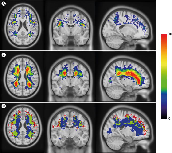
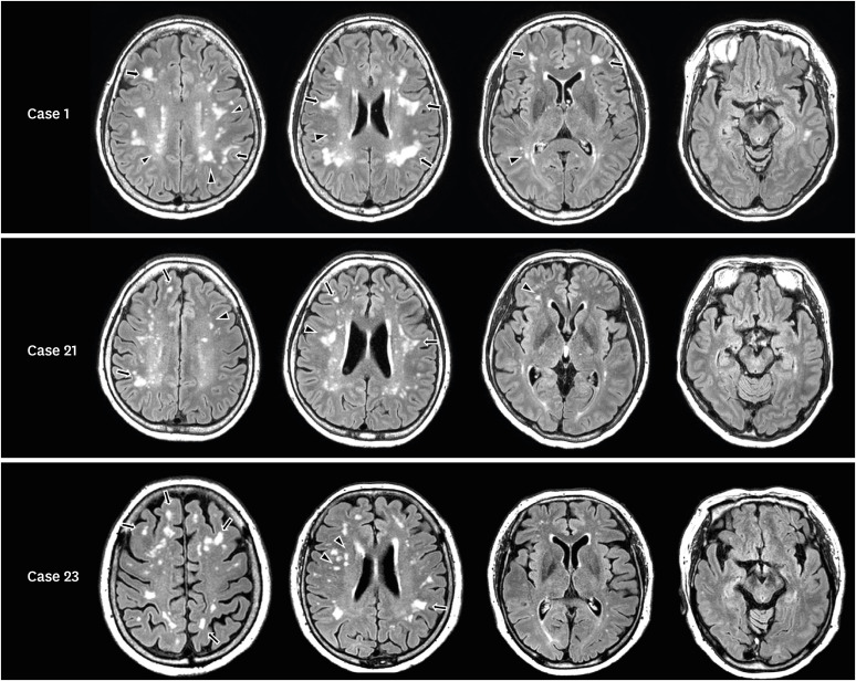
Background and purpose: We investigated the correlation between the deep distribution of white matter hyperintensity (WMH) (dWMH: WMH in deep and corticomedullary areas, with minimal periventricular WMH) and a positive agitated saline contrast echocardiography result.
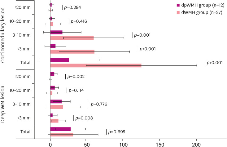
Methods: We retrospectively recruited participants with comprehensive dementia evaluations, an agitated saline study, and brain imaging. The participants were classified into two groups according to WMH-distributions: dWMH and dpWMH (mainly periventricular WMH with or without deep WMH.) We hypothesized that dWMH is more likely associated with embolism, whereas dpWMH is associated with small-vessel diseases. We compared the clinical characteristics, WMH-distributions, and positive rate of agitated saline studies between the two groups.
Results: Among 90 participants, 27 and 12 met the dWMH and dpWMH criteria, respectively. The dWMH-group was younger (62.2±7.5 vs. 78.9±7.3, p<0.001) and had a lower prevalence of hypertension (29.6% vs. 75%, p=0.008), diabetes mellitus (3.7% vs. 25%, p=0.043), and hyperlipidemia (33.3% vs. 83.3%, p=0.043) than the dpWMH-group. Regarding deep white matter lesions, the number of small lesions (<3 mm) was higher in the dWMH-group(10.9±9.7) than in the dpWMH-group (3.1±6.4) (p=0.008), and WMH was predominantly distributed in the border-zones and corticomedullary areas. Most importantly, the positive agitated saline study rate was higher in the dWMH-group than in the dpWMH-group (81.5% vs. 33.3%, p=0.003).
Conclusions: The dWMH-group with younger participants had fewer cardiovascular risk factors, showed more border-zone-distributions, and had a higher agitated saline test positivity rate than the dpWMH-group, indicating that corticomedullary or deep WMH-distribution with minimal periventricular WMH suggests embolic etiologies.

 求助内容:
求助内容: 应助结果提醒方式:


