Matthijs Snelders , Iris H. Koedijk , Julia Schirmer , Otto Mulleners , Juancito van Leeuwen , Nathalie P. de Wagenaar , Oscar Bartulos , Pieter Voskamp , Stefan Braam , Zeno Guttenberg , A.H. Jan Danser , Danielle Majoor-Krakauer , Erik Meijering , Ingrid van der Pluijm , Jeroen Essers
{"title":"使用光学成像分析在硬度可调基质上生长的正常和mybpc3突变的hipsc衍生心肌细胞的收缩压力","authors":"Matthijs Snelders , Iris H. Koedijk , Julia Schirmer , Otto Mulleners , Juancito van Leeuwen , Nathalie P. de Wagenaar , Oscar Bartulos , Pieter Voskamp , Stefan Braam , Zeno Guttenberg , A.H. Jan Danser , Danielle Majoor-Krakauer , Erik Meijering , Ingrid van der Pluijm , Jeroen Essers","doi":"10.1016/j.bbiosy.2022.100068","DOIUrl":null,"url":null,"abstract":"<div><p>Current <em>in vivo</em> disease models and analysis methods for cardiac drug development have been insufficient in providing accurate and reliable predictions of drug efficacy and safety. Here, we propose a custom optical flow-based analysis method to quantitatively measure recordings of contracting cardiomyocytes on polydimethylsiloxane (PDMS), compatible with medium-throughput systems.</p><p>Movement of the PDMS was examined by covalently bound fluorescent beads on the PDMS surface, differences caused by increased substrate stiffness were compared, and cells were stimulated with β-agonist. We further validated the system using cardiomyocytes treated with endothelin-1 and compared their contractions against control and cells incubated with receptor antagonist bosentan. After validation we examined two MYBPC3-mutant patient-derived cell lines.</p><p>Recordings showed that higher substrate stiffness resulted in higher contractile pressure, while beating frequency remained similar to control. β-agonist stimulation resulted in both higher beating frequency as well as higher pressure values during contraction and relaxation. Cells treated with endothelin-1 showed an increased beating frequency, but a lower contraction pressure. Cells treated with both endothelin-1 and bosentan remained at control level of beating frequency and pressure. Lastly, both MYBPC3-mutant lines showed a higher beating frequency and lower contraction pressure.</p><p>Our validated method is capable of automatically quantifying contraction of hiPSC-derived cardiomyocytes on a PDMS substrate of known shear modulus, returning an absolute value. Our method could have major benefits in a medium-throughput setting.</p></div>","PeriodicalId":72379,"journal":{"name":"Biomaterials and biosystems","volume":"8 ","pages":"Article 100068"},"PeriodicalIF":0.0000,"publicationDate":"2022-12-01","publicationTypes":"Journal Article","fieldsOfStudy":null,"isOpenAccess":false,"openAccessPdf":"https://ftp.ncbi.nlm.nih.gov/pub/pmc/oa_pdf/3a/2b/main.PMC9934435.pdf","citationCount":"1","resultStr":"{\"title\":\"Contraction pressure analysis using optical imaging in normal and MYBPC3-mutated hiPSC-derived cardiomyocytes grown on matrices with tunable stiffness\",\"authors\":\"Matthijs Snelders , Iris H. Koedijk , Julia Schirmer , Otto Mulleners , Juancito van Leeuwen , Nathalie P. de Wagenaar , Oscar Bartulos , Pieter Voskamp , Stefan Braam , Zeno Guttenberg , A.H. Jan Danser , Danielle Majoor-Krakauer , Erik Meijering , Ingrid van der Pluijm , Jeroen Essers\",\"doi\":\"10.1016/j.bbiosy.2022.100068\",\"DOIUrl\":null,\"url\":null,\"abstract\":\"<div><p>Current <em>in vivo</em> disease models and analysis methods for cardiac drug development have been insufficient in providing accurate and reliable predictions of drug efficacy and safety. Here, we propose a custom optical flow-based analysis method to quantitatively measure recordings of contracting cardiomyocytes on polydimethylsiloxane (PDMS), compatible with medium-throughput systems.</p><p>Movement of the PDMS was examined by covalently bound fluorescent beads on the PDMS surface, differences caused by increased substrate stiffness were compared, and cells were stimulated with β-agonist. We further validated the system using cardiomyocytes treated with endothelin-1 and compared their contractions against control and cells incubated with receptor antagonist bosentan. After validation we examined two MYBPC3-mutant patient-derived cell lines.</p><p>Recordings showed that higher substrate stiffness resulted in higher contractile pressure, while beating frequency remained similar to control. β-agonist stimulation resulted in both higher beating frequency as well as higher pressure values during contraction and relaxation. Cells treated with endothelin-1 showed an increased beating frequency, but a lower contraction pressure. Cells treated with both endothelin-1 and bosentan remained at control level of beating frequency and pressure. Lastly, both MYBPC3-mutant lines showed a higher beating frequency and lower contraction pressure.</p><p>Our validated method is capable of automatically quantifying contraction of hiPSC-derived cardiomyocytes on a PDMS substrate of known shear modulus, returning an absolute value. Our method could have major benefits in a medium-throughput setting.</p></div>\",\"PeriodicalId\":72379,\"journal\":{\"name\":\"Biomaterials and biosystems\",\"volume\":\"8 \",\"pages\":\"Article 100068\"},\"PeriodicalIF\":0.0000,\"publicationDate\":\"2022-12-01\",\"publicationTypes\":\"Journal Article\",\"fieldsOfStudy\":null,\"isOpenAccess\":false,\"openAccessPdf\":\"https://ftp.ncbi.nlm.nih.gov/pub/pmc/oa_pdf/3a/2b/main.PMC9934435.pdf\",\"citationCount\":\"1\",\"resultStr\":null,\"platform\":\"Semanticscholar\",\"paperid\":null,\"PeriodicalName\":\"Biomaterials and biosystems\",\"FirstCategoryId\":\"1085\",\"ListUrlMain\":\"https://www.sciencedirect.com/science/article/pii/S2666534422000307\",\"RegionNum\":0,\"RegionCategory\":null,\"ArticlePicture\":[],\"TitleCN\":null,\"AbstractTextCN\":null,\"PMCID\":null,\"EPubDate\":\"\",\"PubModel\":\"\",\"JCR\":\"Q3\",\"JCRName\":\"Biochemistry, Genetics and Molecular Biology\",\"Score\":null,\"Total\":0}","platform":"Semanticscholar","paperid":null,"PeriodicalName":"Biomaterials and biosystems","FirstCategoryId":"1085","ListUrlMain":"https://www.sciencedirect.com/science/article/pii/S2666534422000307","RegionNum":0,"RegionCategory":null,"ArticlePicture":[],"TitleCN":null,"AbstractTextCN":null,"PMCID":null,"EPubDate":"","PubModel":"","JCR":"Q3","JCRName":"Biochemistry, Genetics and Molecular Biology","Score":null,"Total":0}
Contraction pressure analysis using optical imaging in normal and MYBPC3-mutated hiPSC-derived cardiomyocytes grown on matrices with tunable stiffness
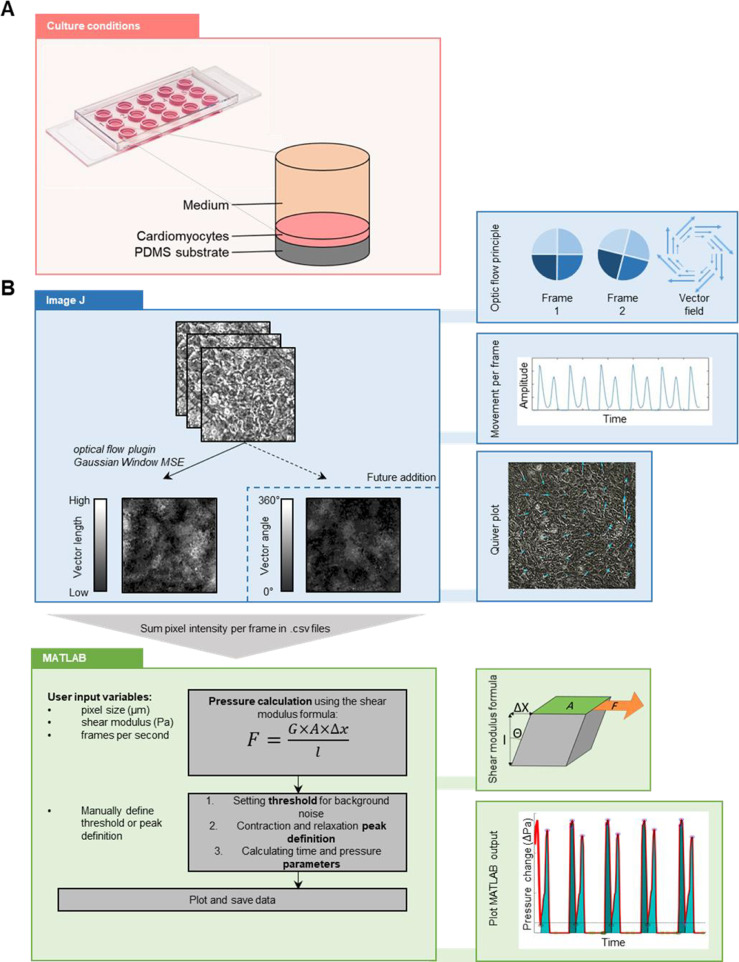
Current in vivo disease models and analysis methods for cardiac drug development have been insufficient in providing accurate and reliable predictions of drug efficacy and safety. Here, we propose a custom optical flow-based analysis method to quantitatively measure recordings of contracting cardiomyocytes on polydimethylsiloxane (PDMS), compatible with medium-throughput systems.
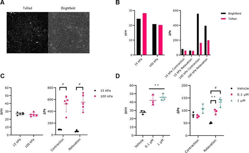
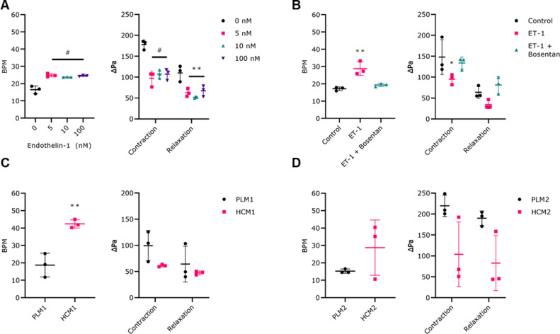
Movement of the PDMS was examined by covalently bound fluorescent beads on the PDMS surface, differences caused by increased substrate stiffness were compared, and cells were stimulated with β-agonist. We further validated the system using cardiomyocytes treated with endothelin-1 and compared their contractions against control and cells incubated with receptor antagonist bosentan. After validation we examined two MYBPC3-mutant patient-derived cell lines.
Recordings showed that higher substrate stiffness resulted in higher contractile pressure, while beating frequency remained similar to control. β-agonist stimulation resulted in both higher beating frequency as well as higher pressure values during contraction and relaxation. Cells treated with endothelin-1 showed an increased beating frequency, but a lower contraction pressure. Cells treated with both endothelin-1 and bosentan remained at control level of beating frequency and pressure. Lastly, both MYBPC3-mutant lines showed a higher beating frequency and lower contraction pressure.
Our validated method is capable of automatically quantifying contraction of hiPSC-derived cardiomyocytes on a PDMS substrate of known shear modulus, returning an absolute value. Our method could have major benefits in a medium-throughput setting.

 求助内容:
求助内容: 应助结果提醒方式:


