{"title":"SynCAM3和细胞周期蛋白D1免疫组化在区分浅表cd34阳性纤维母细胞肿瘤及其组织模拟物中的作用。","authors":"Shintaro Sugita, Tomoko Takenami, Tomomi Kido, Tomoyuki Aoyama, Michiko Hosaka, Keiko Segawa, Taro Sugawara, Hiromi Fujita, Yasutaka Murahashi, Makoto Emori, Atsushi Tsuyuki, Tadashi Hasegawa","doi":"10.1007/s00795-022-00341-w","DOIUrl":null,"url":null,"abstract":"<p><p>Superficial CD34-positive fibroblastic tumor (SCPFT) is a fibroblastic/myofibroblastic soft tissue tumor of rarely metastasizing intermediate malignancy. Some recent studies have described a relationship between SCPFT and PRDM10-rearranged soft tissue tumor (PRT) based on SynCAM3 and PRDM10 expression on immunohistochemistry. We performed CD34, cytokeratin AE1/AE3, SynCAM3, and PRDM10 immunohistochemistry in SCPFT and its histological mimics, including myxoinflammatory fibroblastic sarcoma (MIFS), superficially localized myxofibrosarcoma (MFS), and undifferentiated pleomorphic sarcoma. We also examined cyclin D1 expression because it is expressed in MIFS and MFS. We conducted fluorescence in situ hybridization (FISH) of PRDM10 rearrangement in SCPFT cases. On immunohistochemistry, only SCPFT showed strong and diffuse SynCAM3 expression. SCPFT also exhibited strong nuclear and weak cytoplasmic cyclin D1 expression, which was similar to that observed in MIFS. Two of five SCPFT cases exhibited nuclear PRDM10 expression. FISH revealed PRDM10 split signals in 44% and 24% of tumor cells in two SCPFT cases showing nuclear PRDM10 expression on immunohistochemistry, respectively. A minority of non-SCPFT cases showed focal SynCAM3 expression, but a combination of SynCAM3 and cyclin D1 in addition to CD34 and cytokeratin AE1/AE3 may be useful for the differential diagnosis of SCPFT and its histological mimics.</p>","PeriodicalId":18338,"journal":{"name":"Medical Molecular Morphology","volume":"56 1","pages":"69-77"},"PeriodicalIF":1.2000,"publicationDate":"2023-03-01","publicationTypes":"Journal Article","fieldsOfStudy":null,"isOpenAccess":false,"openAccessPdf":"","citationCount":"2","resultStr":"{\"title\":\"Usefulness of SynCAM3 and cyclin D1 immunohistochemistry in distinguishing superficial CD34-positive fibroblastic tumor from its histological mimics.\",\"authors\":\"Shintaro Sugita, Tomoko Takenami, Tomomi Kido, Tomoyuki Aoyama, Michiko Hosaka, Keiko Segawa, Taro Sugawara, Hiromi Fujita, Yasutaka Murahashi, Makoto Emori, Atsushi Tsuyuki, Tadashi Hasegawa\",\"doi\":\"10.1007/s00795-022-00341-w\",\"DOIUrl\":null,\"url\":null,\"abstract\":\"<p><p>Superficial CD34-positive fibroblastic tumor (SCPFT) is a fibroblastic/myofibroblastic soft tissue tumor of rarely metastasizing intermediate malignancy. Some recent studies have described a relationship between SCPFT and PRDM10-rearranged soft tissue tumor (PRT) based on SynCAM3 and PRDM10 expression on immunohistochemistry. We performed CD34, cytokeratin AE1/AE3, SynCAM3, and PRDM10 immunohistochemistry in SCPFT and its histological mimics, including myxoinflammatory fibroblastic sarcoma (MIFS), superficially localized myxofibrosarcoma (MFS), and undifferentiated pleomorphic sarcoma. We also examined cyclin D1 expression because it is expressed in MIFS and MFS. We conducted fluorescence in situ hybridization (FISH) of PRDM10 rearrangement in SCPFT cases. On immunohistochemistry, only SCPFT showed strong and diffuse SynCAM3 expression. SCPFT also exhibited strong nuclear and weak cytoplasmic cyclin D1 expression, which was similar to that observed in MIFS. Two of five SCPFT cases exhibited nuclear PRDM10 expression. FISH revealed PRDM10 split signals in 44% and 24% of tumor cells in two SCPFT cases showing nuclear PRDM10 expression on immunohistochemistry, respectively. A minority of non-SCPFT cases showed focal SynCAM3 expression, but a combination of SynCAM3 and cyclin D1 in addition to CD34 and cytokeratin AE1/AE3 may be useful for the differential diagnosis of SCPFT and its histological mimics.</p>\",\"PeriodicalId\":18338,\"journal\":{\"name\":\"Medical Molecular Morphology\",\"volume\":\"56 1\",\"pages\":\"69-77\"},\"PeriodicalIF\":1.2000,\"publicationDate\":\"2023-03-01\",\"publicationTypes\":\"Journal Article\",\"fieldsOfStudy\":null,\"isOpenAccess\":false,\"openAccessPdf\":\"\",\"citationCount\":\"2\",\"resultStr\":null,\"platform\":\"Semanticscholar\",\"paperid\":null,\"PeriodicalName\":\"Medical Molecular Morphology\",\"FirstCategoryId\":\"3\",\"ListUrlMain\":\"https://doi.org/10.1007/s00795-022-00341-w\",\"RegionNum\":4,\"RegionCategory\":\"医学\",\"ArticlePicture\":[],\"TitleCN\":null,\"AbstractTextCN\":null,\"PMCID\":null,\"EPubDate\":\"\",\"PubModel\":\"\",\"JCR\":\"Q3\",\"JCRName\":\"PATHOLOGY\",\"Score\":null,\"Total\":0}","platform":"Semanticscholar","paperid":null,"PeriodicalName":"Medical Molecular Morphology","FirstCategoryId":"3","ListUrlMain":"https://doi.org/10.1007/s00795-022-00341-w","RegionNum":4,"RegionCategory":"医学","ArticlePicture":[],"TitleCN":null,"AbstractTextCN":null,"PMCID":null,"EPubDate":"","PubModel":"","JCR":"Q3","JCRName":"PATHOLOGY","Score":null,"Total":0}
Usefulness of SynCAM3 and cyclin D1 immunohistochemistry in distinguishing superficial CD34-positive fibroblastic tumor from its histological mimics.
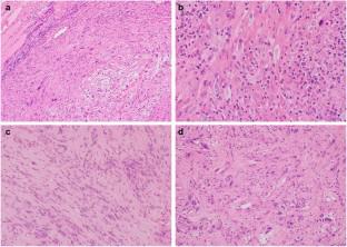
Superficial CD34-positive fibroblastic tumor (SCPFT) is a fibroblastic/myofibroblastic soft tissue tumor of rarely metastasizing intermediate malignancy. Some recent studies have described a relationship between SCPFT and PRDM10-rearranged soft tissue tumor (PRT) based on SynCAM3 and PRDM10 expression on immunohistochemistry. We performed CD34, cytokeratin AE1/AE3, SynCAM3, and PRDM10 immunohistochemistry in SCPFT and its histological mimics, including myxoinflammatory fibroblastic sarcoma (MIFS), superficially localized myxofibrosarcoma (MFS), and undifferentiated pleomorphic sarcoma. We also examined cyclin D1 expression because it is expressed in MIFS and MFS. We conducted fluorescence in situ hybridization (FISH) of PRDM10 rearrangement in SCPFT cases. On immunohistochemistry, only SCPFT showed strong and diffuse SynCAM3 expression. SCPFT also exhibited strong nuclear and weak cytoplasmic cyclin D1 expression, which was similar to that observed in MIFS. Two of five SCPFT cases exhibited nuclear PRDM10 expression. FISH revealed PRDM10 split signals in 44% and 24% of tumor cells in two SCPFT cases showing nuclear PRDM10 expression on immunohistochemistry, respectively. A minority of non-SCPFT cases showed focal SynCAM3 expression, but a combination of SynCAM3 and cyclin D1 in addition to CD34 and cytokeratin AE1/AE3 may be useful for the differential diagnosis of SCPFT and its histological mimics.
期刊介绍:
Medical Molecular Morphology is an international forum for researchers in both basic and clinical medicine to present and discuss new research on the structural mechanisms and the processes of health and disease at the molecular level. The structures of molecules, organelles, cells, tissues, and organs determine their normal function. Disease is thus best understood in terms of structural changes in these different levels of biological organization, especially in molecules and molecular interactions as well as the cellular localization of chemical components. Medical Molecular Morphology welcomes articles on basic or clinical research in the fields of cell biology, molecular biology, and medical, veterinary, and dental sciences using techniques for structural research such as electron microscopy, confocal laser scanning microscopy, enzyme histochemistry, immunohistochemistry, radioautography, X-ray microanalysis, and in situ hybridization.
Manuscripts submitted for publication must contain a statement to the effect that all human studies have been reviewed by the appropriate ethics committee and have therefore been performed in accordance with the ethical standards laid down in an appropriate version of the 1964 Declaration of Helsinki. It should also be stated clearly in the text that all persons gave their informed consent prior to their inclusion in the study. Details that might disclose the identity of the subjects under study should be omitted.
 求助内容:
求助内容: 应助结果提醒方式:


