{"title":"超声诊断乳糜池增大1例。","authors":"Wojciech Łyczek, Bartosz Migda, Michał Kutyłowski","doi":"10.15557/jou.2022.0032","DOIUrl":null,"url":null,"abstract":"<p><strong>Aim of the study: </strong>We present a case report of enlarged cisterna chyli in a 25-year-old woman. The diagnosis was made during a routine abdominal ultrasound examination and afterwards verified with contrast-enhanced MRI.</p><p><strong>Case description: </strong>Ultrasound revealed a large, lobulated, anechoic cystic structure with thin, smooth walls, lacking any solid components. The lesion was located in the retroperitoneal space, beneath the head of the pancreas, between the partially compressed inferior vena cava and the aorta, extending almost to the aortic bifurcation. We performed a contrast-enhanced MRI examination which confirmed the sonographic suspicion of enlarged cisterna chyli, showing a non-enhancing cystic lesion in continuity with the thoracic duct.</p><p><strong>Conclusions: </strong>Anatomy, sonographic and magnetic resonance appearance of cisterna chyli as well as differential diagnosis are discussed.</p>","PeriodicalId":45612,"journal":{"name":"Journal of Ultrasonography","volume":"22 90","pages":"e196-e199"},"PeriodicalIF":1.3000,"publicationDate":"2022-09-01","publicationTypes":"Journal Article","fieldsOfStudy":null,"isOpenAccess":false,"openAccessPdf":"https://ftp.ncbi.nlm.nih.gov/pub/pmc/oa_pdf/13/62/jou-22-e196.PMC9714282.pdf","citationCount":"0","resultStr":"{\"title\":\"Enlarged Cisterna Chyli Diagnosed with Ultrasonography - Case Report.\",\"authors\":\"Wojciech Łyczek, Bartosz Migda, Michał Kutyłowski\",\"doi\":\"10.15557/jou.2022.0032\",\"DOIUrl\":null,\"url\":null,\"abstract\":\"<p><strong>Aim of the study: </strong>We present a case report of enlarged cisterna chyli in a 25-year-old woman. The diagnosis was made during a routine abdominal ultrasound examination and afterwards verified with contrast-enhanced MRI.</p><p><strong>Case description: </strong>Ultrasound revealed a large, lobulated, anechoic cystic structure with thin, smooth walls, lacking any solid components. The lesion was located in the retroperitoneal space, beneath the head of the pancreas, between the partially compressed inferior vena cava and the aorta, extending almost to the aortic bifurcation. We performed a contrast-enhanced MRI examination which confirmed the sonographic suspicion of enlarged cisterna chyli, showing a non-enhancing cystic lesion in continuity with the thoracic duct.</p><p><strong>Conclusions: </strong>Anatomy, sonographic and magnetic resonance appearance of cisterna chyli as well as differential diagnosis are discussed.</p>\",\"PeriodicalId\":45612,\"journal\":{\"name\":\"Journal of Ultrasonography\",\"volume\":\"22 90\",\"pages\":\"e196-e199\"},\"PeriodicalIF\":1.3000,\"publicationDate\":\"2022-09-01\",\"publicationTypes\":\"Journal Article\",\"fieldsOfStudy\":null,\"isOpenAccess\":false,\"openAccessPdf\":\"https://ftp.ncbi.nlm.nih.gov/pub/pmc/oa_pdf/13/62/jou-22-e196.PMC9714282.pdf\",\"citationCount\":\"0\",\"resultStr\":null,\"platform\":\"Semanticscholar\",\"paperid\":null,\"PeriodicalName\":\"Journal of Ultrasonography\",\"FirstCategoryId\":\"1085\",\"ListUrlMain\":\"https://doi.org/10.15557/jou.2022.0032\",\"RegionNum\":0,\"RegionCategory\":null,\"ArticlePicture\":[],\"TitleCN\":null,\"AbstractTextCN\":null,\"PMCID\":null,\"EPubDate\":\"\",\"PubModel\":\"\",\"JCR\":\"Q3\",\"JCRName\":\"RADIOLOGY, NUCLEAR MEDICINE & MEDICAL IMAGING\",\"Score\":null,\"Total\":0}","platform":"Semanticscholar","paperid":null,"PeriodicalName":"Journal of Ultrasonography","FirstCategoryId":"1085","ListUrlMain":"https://doi.org/10.15557/jou.2022.0032","RegionNum":0,"RegionCategory":null,"ArticlePicture":[],"TitleCN":null,"AbstractTextCN":null,"PMCID":null,"EPubDate":"","PubModel":"","JCR":"Q3","JCRName":"RADIOLOGY, NUCLEAR MEDICINE & MEDICAL IMAGING","Score":null,"Total":0}
Enlarged Cisterna Chyli Diagnosed with Ultrasonography - Case Report.
Aim of the study: We present a case report of enlarged cisterna chyli in a 25-year-old woman. The diagnosis was made during a routine abdominal ultrasound examination and afterwards verified with contrast-enhanced MRI.
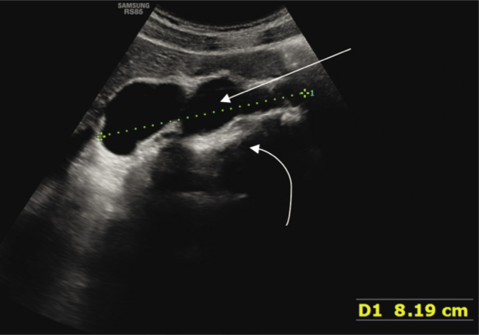
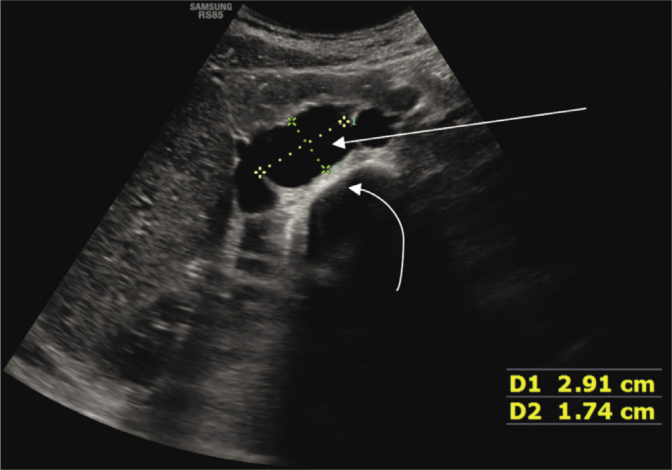
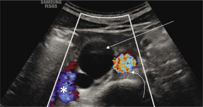
Case description: Ultrasound revealed a large, lobulated, anechoic cystic structure with thin, smooth walls, lacking any solid components. The lesion was located in the retroperitoneal space, beneath the head of the pancreas, between the partially compressed inferior vena cava and the aorta, extending almost to the aortic bifurcation. We performed a contrast-enhanced MRI examination which confirmed the sonographic suspicion of enlarged cisterna chyli, showing a non-enhancing cystic lesion in continuity with the thoracic duct.
Conclusions: Anatomy, sonographic and magnetic resonance appearance of cisterna chyli as well as differential diagnosis are discussed.

 求助内容:
求助内容: 应助结果提醒方式:


