Salah M Fateh, Lusan A Arkawazi, Soran H Tahir, Rezheen J Rashid, Dalshad H Rahman, Ismaeel Aghaways, Fahmi H Kakamad, Abdulwahid M Salih, Rawa Bapir, Saman S Fakhralddin, Fattah H Fattah, Berun A Abdalla, Shvan H Mohammed
{"title":"肾细胞癌T分期:术前增强计算机断层扫描的诊断准确性。","authors":"Salah M Fateh, Lusan A Arkawazi, Soran H Tahir, Rezheen J Rashid, Dalshad H Rahman, Ismaeel Aghaways, Fahmi H Kakamad, Abdulwahid M Salih, Rawa Bapir, Saman S Fakhralddin, Fattah H Fattah, Berun A Abdalla, Shvan H Mohammed","doi":"10.3892/mco.2023.2607","DOIUrl":null,"url":null,"abstract":"<p><p>Renal cell carcinoma (RCC) accounts for 1-2% of all malignancies and is the most common renal tumor in adults. Imaging studies are used for diagnosis and staging. Tumor-Node-Metastasis staging strongly affects prognosis and management, while contrast-enhanced computed tomography (CECT) is regarded as a standard imaging technique for local and distant staging. The present study aimed to evaluate the accuracy of CECT for the preoperative staging of RCC by using surgical and pathological staging as the reference methods. This single-center prospective study was conducted between October 2019 and November 2021. The preoperative abdominal CT scans of patients suspected of having RCC were reviewed. Imaging data were collected, including tumor side and size, and perinephric fat invasion. Intraoperative notes were recorded, including the operation type, perinephric fat invasion, renal vein (RV) or inferior vena cava (IVC) tumor extension, and surrounding organ invasion. pathological data were collected on tumor size, RCC type, presence of clear margins, presence of renal capsule or perinephric fat invasion, renal sinus or pelvicalyceal system (PCS) invasion, segmental or main RV extension, and the involvement of Gerota's fascia and nearby organs. Preoperative CECT revealed that 42 out of 59 tumors had a greater maximum diameter than the pathological specimen, with an overall disparity of 0.25 cm. The specificity of CT for the detection of tumor invasion of the perinephric and renal sinus fat and PCS was 95%, and the sensitivity ranged from 80 to 88%. CT had an 83% sensitivity and a 95 specificity in detecting T4 stage cancer, with a 100% specificity for adrenal invasion. The concordance between radiographic and histological results for RV and IVC involvement was high, with specificities of 94 and 98%, and sensitivities of 80 and 100%, respectively. Overall accuracy for correct T staging was 80%. In conclusion, CECT is accurate in the local T staging of RCC, with high sensitivity and specificity for estimating tumor size and detecting extension to nearby structures and venous invasion.</p>","PeriodicalId":18737,"journal":{"name":"Molecular and clinical oncology","volume":"18 2","pages":"11"},"PeriodicalIF":1.4000,"publicationDate":"2023-02-01","publicationTypes":"Journal Article","fieldsOfStudy":null,"isOpenAccess":false,"openAccessPdf":"https://ftp.ncbi.nlm.nih.gov/pub/pmc/oa_pdf/6e/3f/mco-18-02-02607.PMC9892965.pdf","citationCount":"1","resultStr":"{\"title\":\"Renal cell carcinoma T staging: Diagnostic accuracy of preoperative contrast-enhanced computed tomography.\",\"authors\":\"Salah M Fateh, Lusan A Arkawazi, Soran H Tahir, Rezheen J Rashid, Dalshad H Rahman, Ismaeel Aghaways, Fahmi H Kakamad, Abdulwahid M Salih, Rawa Bapir, Saman S Fakhralddin, Fattah H Fattah, Berun A Abdalla, Shvan H Mohammed\",\"doi\":\"10.3892/mco.2023.2607\",\"DOIUrl\":null,\"url\":null,\"abstract\":\"<p><p>Renal cell carcinoma (RCC) accounts for 1-2% of all malignancies and is the most common renal tumor in adults. Imaging studies are used for diagnosis and staging. Tumor-Node-Metastasis staging strongly affects prognosis and management, while contrast-enhanced computed tomography (CECT) is regarded as a standard imaging technique for local and distant staging. The present study aimed to evaluate the accuracy of CECT for the preoperative staging of RCC by using surgical and pathological staging as the reference methods. This single-center prospective study was conducted between October 2019 and November 2021. The preoperative abdominal CT scans of patients suspected of having RCC were reviewed. Imaging data were collected, including tumor side and size, and perinephric fat invasion. Intraoperative notes were recorded, including the operation type, perinephric fat invasion, renal vein (RV) or inferior vena cava (IVC) tumor extension, and surrounding organ invasion. pathological data were collected on tumor size, RCC type, presence of clear margins, presence of renal capsule or perinephric fat invasion, renal sinus or pelvicalyceal system (PCS) invasion, segmental or main RV extension, and the involvement of Gerota's fascia and nearby organs. Preoperative CECT revealed that 42 out of 59 tumors had a greater maximum diameter than the pathological specimen, with an overall disparity of 0.25 cm. The specificity of CT for the detection of tumor invasion of the perinephric and renal sinus fat and PCS was 95%, and the sensitivity ranged from 80 to 88%. CT had an 83% sensitivity and a 95 specificity in detecting T4 stage cancer, with a 100% specificity for adrenal invasion. The concordance between radiographic and histological results for RV and IVC involvement was high, with specificities of 94 and 98%, and sensitivities of 80 and 100%, respectively. Overall accuracy for correct T staging was 80%. In conclusion, CECT is accurate in the local T staging of RCC, with high sensitivity and specificity for estimating tumor size and detecting extension to nearby structures and venous invasion.</p>\",\"PeriodicalId\":18737,\"journal\":{\"name\":\"Molecular and clinical oncology\",\"volume\":\"18 2\",\"pages\":\"11\"},\"PeriodicalIF\":1.4000,\"publicationDate\":\"2023-02-01\",\"publicationTypes\":\"Journal Article\",\"fieldsOfStudy\":null,\"isOpenAccess\":false,\"openAccessPdf\":\"https://ftp.ncbi.nlm.nih.gov/pub/pmc/oa_pdf/6e/3f/mco-18-02-02607.PMC9892965.pdf\",\"citationCount\":\"1\",\"resultStr\":null,\"platform\":\"Semanticscholar\",\"paperid\":null,\"PeriodicalName\":\"Molecular and clinical oncology\",\"FirstCategoryId\":\"1085\",\"ListUrlMain\":\"https://doi.org/10.3892/mco.2023.2607\",\"RegionNum\":0,\"RegionCategory\":null,\"ArticlePicture\":[],\"TitleCN\":null,\"AbstractTextCN\":null,\"PMCID\":null,\"EPubDate\":\"\",\"PubModel\":\"\",\"JCR\":\"Q4\",\"JCRName\":\"ONCOLOGY\",\"Score\":null,\"Total\":0}","platform":"Semanticscholar","paperid":null,"PeriodicalName":"Molecular and clinical oncology","FirstCategoryId":"1085","ListUrlMain":"https://doi.org/10.3892/mco.2023.2607","RegionNum":0,"RegionCategory":null,"ArticlePicture":[],"TitleCN":null,"AbstractTextCN":null,"PMCID":null,"EPubDate":"","PubModel":"","JCR":"Q4","JCRName":"ONCOLOGY","Score":null,"Total":0}
Renal cell carcinoma T staging: Diagnostic accuracy of preoperative contrast-enhanced computed tomography.
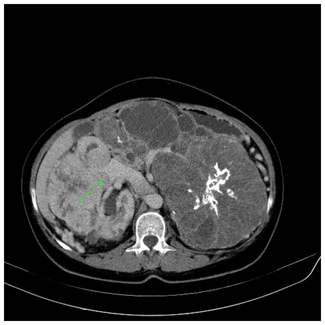
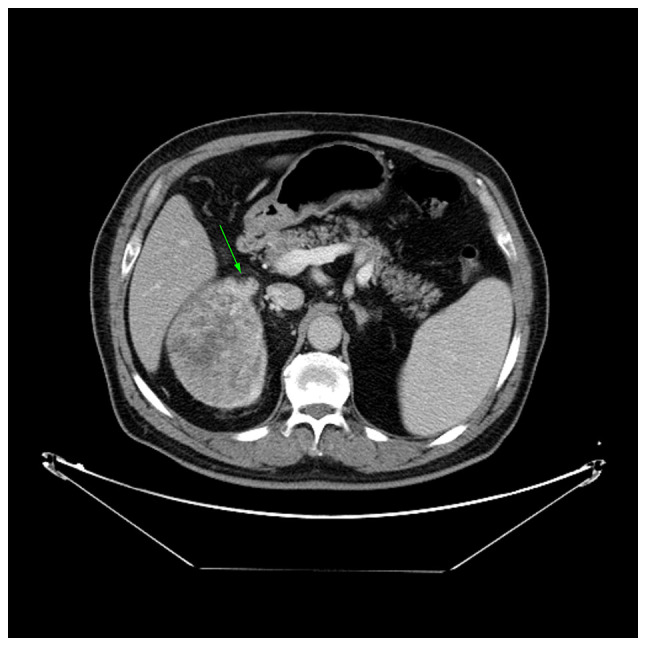
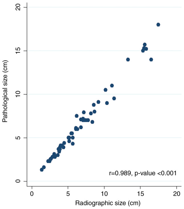
Renal cell carcinoma (RCC) accounts for 1-2% of all malignancies and is the most common renal tumor in adults. Imaging studies are used for diagnosis and staging. Tumor-Node-Metastasis staging strongly affects prognosis and management, while contrast-enhanced computed tomography (CECT) is regarded as a standard imaging technique for local and distant staging. The present study aimed to evaluate the accuracy of CECT for the preoperative staging of RCC by using surgical and pathological staging as the reference methods. This single-center prospective study was conducted between October 2019 and November 2021. The preoperative abdominal CT scans of patients suspected of having RCC were reviewed. Imaging data were collected, including tumor side and size, and perinephric fat invasion. Intraoperative notes were recorded, including the operation type, perinephric fat invasion, renal vein (RV) or inferior vena cava (IVC) tumor extension, and surrounding organ invasion. pathological data were collected on tumor size, RCC type, presence of clear margins, presence of renal capsule or perinephric fat invasion, renal sinus or pelvicalyceal system (PCS) invasion, segmental or main RV extension, and the involvement of Gerota's fascia and nearby organs. Preoperative CECT revealed that 42 out of 59 tumors had a greater maximum diameter than the pathological specimen, with an overall disparity of 0.25 cm. The specificity of CT for the detection of tumor invasion of the perinephric and renal sinus fat and PCS was 95%, and the sensitivity ranged from 80 to 88%. CT had an 83% sensitivity and a 95 specificity in detecting T4 stage cancer, with a 100% specificity for adrenal invasion. The concordance between radiographic and histological results for RV and IVC involvement was high, with specificities of 94 and 98%, and sensitivities of 80 and 100%, respectively. Overall accuracy for correct T staging was 80%. In conclusion, CECT is accurate in the local T staging of RCC, with high sensitivity and specificity for estimating tumor size and detecting extension to nearby structures and venous invasion.

 求助内容:
求助内容: 应助结果提醒方式:


