Tin Hoang Nguyen, Kien Trung Nguyen, Long Duc Tran, An Thi Thuy Le, Thu Minh Phung, Truc Thi Ngoc Banh, Trang Thi Vo, Michael Bodo
{"title":"绝经期妇女血流脑电图特征及相关因素分析。","authors":"Tin Hoang Nguyen, Kien Trung Nguyen, Long Duc Tran, An Thi Thuy Le, Thu Minh Phung, Truc Thi Ngoc Banh, Trang Thi Vo, Michael Bodo","doi":"10.2478/joeb-2022-0012","DOIUrl":null,"url":null,"abstract":"<p><p>The significant drop in estrogen levels during menopause increases the cardiovascular risks, one of which is cerebrovascular atherosclerosis. Research on rheoencephalography (REG) parameters for the early diagnosis of cerebrovascular atherosclerotic lesions is of great interest to scientists because of its ease of implementation, low cost, and non-invasiveness. The objectives of study are to evaluate the vascular tone, cerebral circulation flow in each hemisphere of the brain of menopausal women, and some associated factors through waveform characteristics and parameters in REG. A controlled cross-sectional descriptive study was conducted on a group of patients including 80 menopausal women and a control group of 46 menstruating women. All patients were measured REG in the frontal-occipital leads by VasoScreen 5000 impedance REG meter. In menopausal women, the percentage of sharp waves, the percentage of clear side waves, and the average REG were all lower than in the control group (p<0.01). The mean conduction time and mean slope ratio was lower than the control group (p<0.001). The mean peak time was higher than the control group (p<0.01). The mean elasticity index (alpha/T) was higher than the control group (p<0.001). Menopausal women have increased vascular tone, the highest in the group of women 50-60 years old, menopause <5 years, having a habit of eating red meat; and decreased blood flow intensity, the highest in the group of women <50 years old. However, the difference was statistically significant only in the left hemisphere (p<0.05). Vascular hypertonia in menopausal women with central obesity was higher than in the non-obese group in both hemispheres (p<0.05). In conclusion, menopausal women had atherosclerosis in both hemispheres of the brain, which was clearly shown in the rate of increased vascular tone. Central obesity may increase the risk of vascular hypertonia 3.75 times in the right and 5.44 times in the left hemisphere.</p>","PeriodicalId":38125,"journal":{"name":"Journal of Electrical Bioimpedance","volume":"13 1","pages":"78-87"},"PeriodicalIF":0.0000,"publicationDate":"2022-01-01","publicationTypes":"Journal Article","fieldsOfStudy":null,"isOpenAccess":false,"openAccessPdf":"https://www.ncbi.nlm.nih.gov/pmc/articles/PMC9837873/pdf/","citationCount":"0","resultStr":"{\"title\":\"Characteristics of Rheoencephalography and some Associated Factors on Menopausal Women.\",\"authors\":\"Tin Hoang Nguyen, Kien Trung Nguyen, Long Duc Tran, An Thi Thuy Le, Thu Minh Phung, Truc Thi Ngoc Banh, Trang Thi Vo, Michael Bodo\",\"doi\":\"10.2478/joeb-2022-0012\",\"DOIUrl\":null,\"url\":null,\"abstract\":\"<p><p>The significant drop in estrogen levels during menopause increases the cardiovascular risks, one of which is cerebrovascular atherosclerosis. Research on rheoencephalography (REG) parameters for the early diagnosis of cerebrovascular atherosclerotic lesions is of great interest to scientists because of its ease of implementation, low cost, and non-invasiveness. The objectives of study are to evaluate the vascular tone, cerebral circulation flow in each hemisphere of the brain of menopausal women, and some associated factors through waveform characteristics and parameters in REG. A controlled cross-sectional descriptive study was conducted on a group of patients including 80 menopausal women and a control group of 46 menstruating women. All patients were measured REG in the frontal-occipital leads by VasoScreen 5000 impedance REG meter. In menopausal women, the percentage of sharp waves, the percentage of clear side waves, and the average REG were all lower than in the control group (p<0.01). The mean conduction time and mean slope ratio was lower than the control group (p<0.001). The mean peak time was higher than the control group (p<0.01). The mean elasticity index (alpha/T) was higher than the control group (p<0.001). Menopausal women have increased vascular tone, the highest in the group of women 50-60 years old, menopause <5 years, having a habit of eating red meat; and decreased blood flow intensity, the highest in the group of women <50 years old. However, the difference was statistically significant only in the left hemisphere (p<0.05). Vascular hypertonia in menopausal women with central obesity was higher than in the non-obese group in both hemispheres (p<0.05). In conclusion, menopausal women had atherosclerosis in both hemispheres of the brain, which was clearly shown in the rate of increased vascular tone. Central obesity may increase the risk of vascular hypertonia 3.75 times in the right and 5.44 times in the left hemisphere.</p>\",\"PeriodicalId\":38125,\"journal\":{\"name\":\"Journal of Electrical Bioimpedance\",\"volume\":\"13 1\",\"pages\":\"78-87\"},\"PeriodicalIF\":0.0000,\"publicationDate\":\"2022-01-01\",\"publicationTypes\":\"Journal Article\",\"fieldsOfStudy\":null,\"isOpenAccess\":false,\"openAccessPdf\":\"https://www.ncbi.nlm.nih.gov/pmc/articles/PMC9837873/pdf/\",\"citationCount\":\"0\",\"resultStr\":null,\"platform\":\"Semanticscholar\",\"paperid\":null,\"PeriodicalName\":\"Journal of Electrical Bioimpedance\",\"FirstCategoryId\":\"1085\",\"ListUrlMain\":\"https://doi.org/10.2478/joeb-2022-0012\",\"RegionNum\":0,\"RegionCategory\":null,\"ArticlePicture\":[],\"TitleCN\":null,\"AbstractTextCN\":null,\"PMCID\":null,\"EPubDate\":\"\",\"PubModel\":\"\",\"JCR\":\"Q3\",\"JCRName\":\"Biochemistry, Genetics and Molecular Biology\",\"Score\":null,\"Total\":0}","platform":"Semanticscholar","paperid":null,"PeriodicalName":"Journal of Electrical Bioimpedance","FirstCategoryId":"1085","ListUrlMain":"https://doi.org/10.2478/joeb-2022-0012","RegionNum":0,"RegionCategory":null,"ArticlePicture":[],"TitleCN":null,"AbstractTextCN":null,"PMCID":null,"EPubDate":"","PubModel":"","JCR":"Q3","JCRName":"Biochemistry, Genetics and Molecular Biology","Score":null,"Total":0}
Characteristics of Rheoencephalography and some Associated Factors on Menopausal Women.
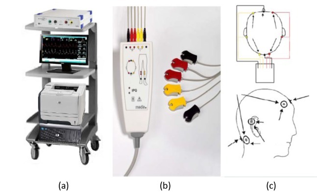
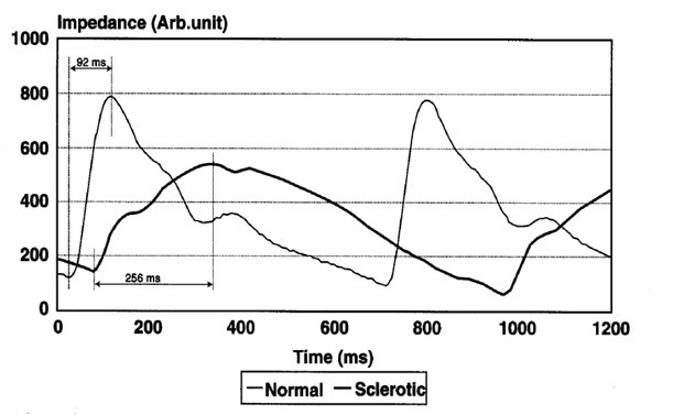
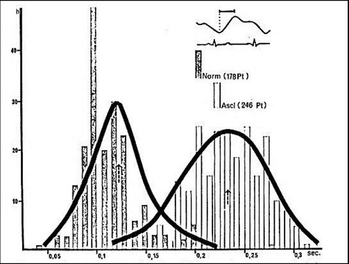
The significant drop in estrogen levels during menopause increases the cardiovascular risks, one of which is cerebrovascular atherosclerosis. Research on rheoencephalography (REG) parameters for the early diagnosis of cerebrovascular atherosclerotic lesions is of great interest to scientists because of its ease of implementation, low cost, and non-invasiveness. The objectives of study are to evaluate the vascular tone, cerebral circulation flow in each hemisphere of the brain of menopausal women, and some associated factors through waveform characteristics and parameters in REG. A controlled cross-sectional descriptive study was conducted on a group of patients including 80 menopausal women and a control group of 46 menstruating women. All patients were measured REG in the frontal-occipital leads by VasoScreen 5000 impedance REG meter. In menopausal women, the percentage of sharp waves, the percentage of clear side waves, and the average REG were all lower than in the control group (p<0.01). The mean conduction time and mean slope ratio was lower than the control group (p<0.001). The mean peak time was higher than the control group (p<0.01). The mean elasticity index (alpha/T) was higher than the control group (p<0.001). Menopausal women have increased vascular tone, the highest in the group of women 50-60 years old, menopause <5 years, having a habit of eating red meat; and decreased blood flow intensity, the highest in the group of women <50 years old. However, the difference was statistically significant only in the left hemisphere (p<0.05). Vascular hypertonia in menopausal women with central obesity was higher than in the non-obese group in both hemispheres (p<0.05). In conclusion, menopausal women had atherosclerosis in both hemispheres of the brain, which was clearly shown in the rate of increased vascular tone. Central obesity may increase the risk of vascular hypertonia 3.75 times in the right and 5.44 times in the left hemisphere.

 求助内容:
求助内容: 应助结果提醒方式:


