Se-Jung Yoon, Sungha Park, Eui-Young Choi, Hye-Sun Seo, Chi Young Shim, Chul Min Ahn, Sung-Ai Kim, Jong-Won Ha
{"title":"左房速度矢量成像评价左室肥厚和肥厚型心肌病早期舒张功能障碍。","authors":"Se-Jung Yoon, Sungha Park, Eui-Young Choi, Hye-Sun Seo, Chi Young Shim, Chul Min Ahn, Sung-Ai Kim, Jong-Won Ha","doi":"10.4250/jcvi.2022.0064","DOIUrl":null,"url":null,"abstract":"<p><strong>Background: </strong>The function of left atrium (LA) is difficult to assess because of its ventricle-dependent, dynamic movement. The aim of this study was to assess LA function using velocity vector imaging (VVI) and compare LA function in patients with hypertrophic cardiomyopathy (HCMP) and left ventricular hypertrophy (LVH) with normal controls.</p><p><strong>Methods: </strong>Fourteen patients with HCMP (72% male, mean age of 52.6 ± 9.8), 15 hypertensive patients with LVH (88% male, mean age of 54.0 ± 15.3), and 10 age-matched controls (83% male, mean age of 50.0 ± 4.6) were prospectively studied. Echocardiographic images of the LA were analyzed with VVI, and strain rate (SR) was compared among the 3 groups.</p><p><strong>Results: </strong>The e' velocity (7.7 ± 1.1; 5.1 ± 0.8; 4.5 ± 1.3 cm/sec, p = 0.013), E/e' (6.8 ± 1.6; 12.4 ± 3.3; 14.7 ± 4.2, p = 0.035), and late diastolic SR at mid LA (-1.65 ± 0.51; -0.97 ± 0.55; -0.82 ± 0.32, p = 0.002) were significantly different among the groups (normal; LVH; HCMP, respectively). The e' velocity, E/e', and late diastolic SR at mid LA were significantly different between normal and LVH (p = 0.001; 0.022; 0.018), whereas LA size was similar between normal and LVH (p = 0.592). The mean late diastolic peak SR of mid LA was significantly correlated with indices of diastolic function (E/e', e', and LA size).</p><p><strong>Conclusions: </strong>The SR is a useful tool for detailed evaluation of LA function, especially early dysfunction of LA in groups with normal LA size.</p>","PeriodicalId":15229,"journal":{"name":"Journal of Cardiovascular Imaging","volume":"31 1","pages":"41-48"},"PeriodicalIF":0.0000,"publicationDate":"2023-01-01","publicationTypes":"Journal Article","fieldsOfStudy":null,"isOpenAccess":false,"openAccessPdf":"https://ftp.ncbi.nlm.nih.gov/pub/pmc/oa_pdf/0c/44/jcvi-31-41.PMC9880349.pdf","citationCount":"3","resultStr":"{\"title\":\"Left Atrial Velocity Vector Imaging Can Assess Early Diastolic Dysfunction in Left Ventricular Hypertrophy and Hypertrophic Cardiomyopathy.\",\"authors\":\"Se-Jung Yoon, Sungha Park, Eui-Young Choi, Hye-Sun Seo, Chi Young Shim, Chul Min Ahn, Sung-Ai Kim, Jong-Won Ha\",\"doi\":\"10.4250/jcvi.2022.0064\",\"DOIUrl\":null,\"url\":null,\"abstract\":\"<p><strong>Background: </strong>The function of left atrium (LA) is difficult to assess because of its ventricle-dependent, dynamic movement. The aim of this study was to assess LA function using velocity vector imaging (VVI) and compare LA function in patients with hypertrophic cardiomyopathy (HCMP) and left ventricular hypertrophy (LVH) with normal controls.</p><p><strong>Methods: </strong>Fourteen patients with HCMP (72% male, mean age of 52.6 ± 9.8), 15 hypertensive patients with LVH (88% male, mean age of 54.0 ± 15.3), and 10 age-matched controls (83% male, mean age of 50.0 ± 4.6) were prospectively studied. Echocardiographic images of the LA were analyzed with VVI, and strain rate (SR) was compared among the 3 groups.</p><p><strong>Results: </strong>The e' velocity (7.7 ± 1.1; 5.1 ± 0.8; 4.5 ± 1.3 cm/sec, p = 0.013), E/e' (6.8 ± 1.6; 12.4 ± 3.3; 14.7 ± 4.2, p = 0.035), and late diastolic SR at mid LA (-1.65 ± 0.51; -0.97 ± 0.55; -0.82 ± 0.32, p = 0.002) were significantly different among the groups (normal; LVH; HCMP, respectively). The e' velocity, E/e', and late diastolic SR at mid LA were significantly different between normal and LVH (p = 0.001; 0.022; 0.018), whereas LA size was similar between normal and LVH (p = 0.592). The mean late diastolic peak SR of mid LA was significantly correlated with indices of diastolic function (E/e', e', and LA size).</p><p><strong>Conclusions: </strong>The SR is a useful tool for detailed evaluation of LA function, especially early dysfunction of LA in groups with normal LA size.</p>\",\"PeriodicalId\":15229,\"journal\":{\"name\":\"Journal of Cardiovascular Imaging\",\"volume\":\"31 1\",\"pages\":\"41-48\"},\"PeriodicalIF\":0.0000,\"publicationDate\":\"2023-01-01\",\"publicationTypes\":\"Journal Article\",\"fieldsOfStudy\":null,\"isOpenAccess\":false,\"openAccessPdf\":\"https://ftp.ncbi.nlm.nih.gov/pub/pmc/oa_pdf/0c/44/jcvi-31-41.PMC9880349.pdf\",\"citationCount\":\"3\",\"resultStr\":null,\"platform\":\"Semanticscholar\",\"paperid\":null,\"PeriodicalName\":\"Journal of Cardiovascular Imaging\",\"FirstCategoryId\":\"1085\",\"ListUrlMain\":\"https://doi.org/10.4250/jcvi.2022.0064\",\"RegionNum\":0,\"RegionCategory\":null,\"ArticlePicture\":[],\"TitleCN\":null,\"AbstractTextCN\":null,\"PMCID\":null,\"EPubDate\":\"\",\"PubModel\":\"\",\"JCR\":\"Q2\",\"JCRName\":\"Medicine\",\"Score\":null,\"Total\":0}","platform":"Semanticscholar","paperid":null,"PeriodicalName":"Journal of Cardiovascular Imaging","FirstCategoryId":"1085","ListUrlMain":"https://doi.org/10.4250/jcvi.2022.0064","RegionNum":0,"RegionCategory":null,"ArticlePicture":[],"TitleCN":null,"AbstractTextCN":null,"PMCID":null,"EPubDate":"","PubModel":"","JCR":"Q2","JCRName":"Medicine","Score":null,"Total":0}
Left Atrial Velocity Vector Imaging Can Assess Early Diastolic Dysfunction in Left Ventricular Hypertrophy and Hypertrophic Cardiomyopathy.
Background: The function of left atrium (LA) is difficult to assess because of its ventricle-dependent, dynamic movement. The aim of this study was to assess LA function using velocity vector imaging (VVI) and compare LA function in patients with hypertrophic cardiomyopathy (HCMP) and left ventricular hypertrophy (LVH) with normal controls.
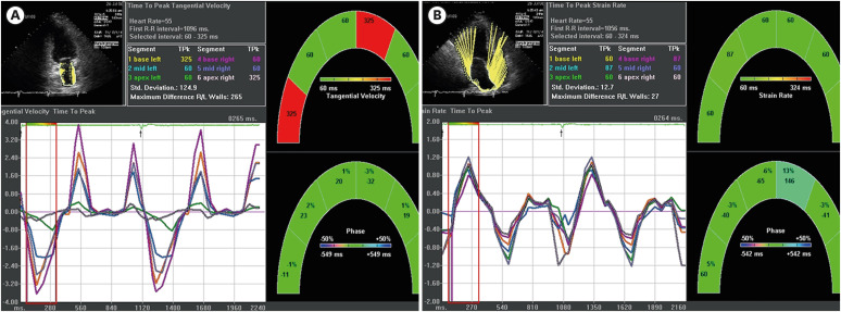
Methods: Fourteen patients with HCMP (72% male, mean age of 52.6 ± 9.8), 15 hypertensive patients with LVH (88% male, mean age of 54.0 ± 15.3), and 10 age-matched controls (83% male, mean age of 50.0 ± 4.6) were prospectively studied. Echocardiographic images of the LA were analyzed with VVI, and strain rate (SR) was compared among the 3 groups.
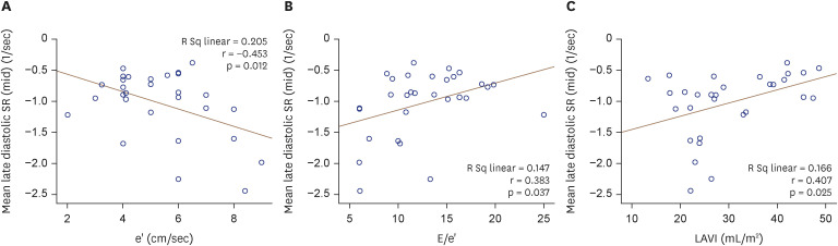
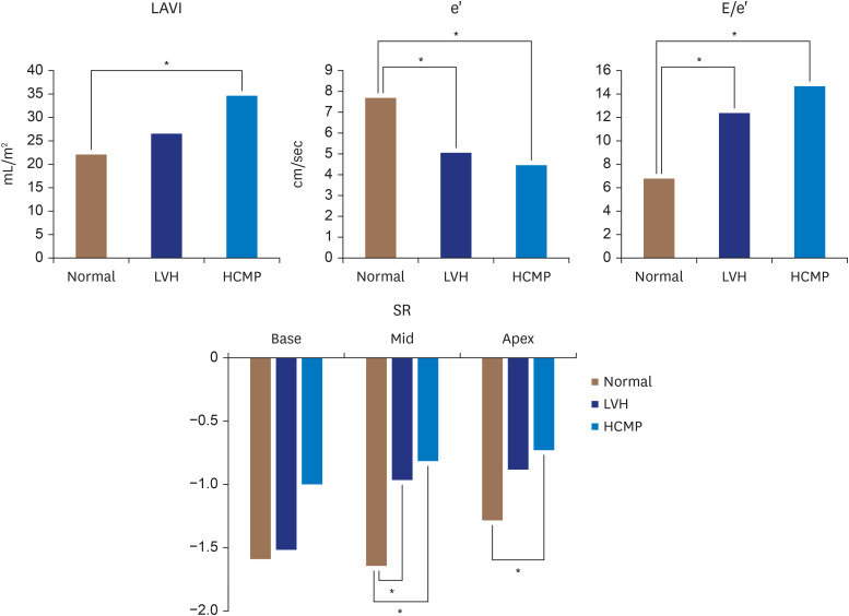
Results: The e' velocity (7.7 ± 1.1; 5.1 ± 0.8; 4.5 ± 1.3 cm/sec, p = 0.013), E/e' (6.8 ± 1.6; 12.4 ± 3.3; 14.7 ± 4.2, p = 0.035), and late diastolic SR at mid LA (-1.65 ± 0.51; -0.97 ± 0.55; -0.82 ± 0.32, p = 0.002) were significantly different among the groups (normal; LVH; HCMP, respectively). The e' velocity, E/e', and late diastolic SR at mid LA were significantly different between normal and LVH (p = 0.001; 0.022; 0.018), whereas LA size was similar between normal and LVH (p = 0.592). The mean late diastolic peak SR of mid LA was significantly correlated with indices of diastolic function (E/e', e', and LA size).
Conclusions: The SR is a useful tool for detailed evaluation of LA function, especially early dysfunction of LA in groups with normal LA size.

 求助内容:
求助内容: 应助结果提醒方式:


