{"title":"偏振与非偏振数字图像测量牙龈黑色素沉着的效度和信度。","authors":"Talal M Zahid, Zuhair S Natto","doi":"10.2147/CCIDE.S422139","DOIUrl":null,"url":null,"abstract":"<p><strong>Aim: </strong>This study aimed to compare the validity and reliability of polarized and non-polarized intraoral photography for the measurement of gingival melanin pigmentation.</p><p><strong>Materials and methods: </strong>A case series study was conducted on ten patients scheduled for gingival depigmentation. A total of 976 polarized and non-polarized image samples were collected, capturing two rows above the gingival margin, for analysis. These images were taken both before and one year after the depigmentation procedure. Three independent evaluators assessed the photographs (an orthodontist, a general dentist, and a layperson). The Dummett Oral Pigmentation Index (DOPI) and Gingival Melanosis Record (GMR) indices were used to measure the level of gingival pigmentation.</p><p><strong>Results: </strong>The study found no significant differences between polarized and non-polarized images taken before and after depigmentation. Both methods of imaging received similar scores from the evaluators. The orthodontist identified more pigmented slides than the layperson and the general dentist.</p><p><strong>Conclusion: </strong>Both polarized and non-polarized photographic methods may be used for assessing gingival pigmentation. However, further research is warranted to confirm this finding and examine additional factors.</p>","PeriodicalId":10445,"journal":{"name":"Clinical, Cosmetic and Investigational Dentistry","volume":"15 ","pages":"189-197"},"PeriodicalIF":1.5000,"publicationDate":"2023-01-01","publicationTypes":"Journal Article","fieldsOfStudy":null,"isOpenAccess":false,"openAccessPdf":"https://ftp.ncbi.nlm.nih.gov/pub/pmc/oa_pdf/08/d7/ccide-15-189.PMC10504902.pdf","citationCount":"0","resultStr":"{\"title\":\"Validity and Reliability of Polarized vs Non-Polarized Digital Images for Measuring Gingival Melanin Pigmentation.\",\"authors\":\"Talal M Zahid, Zuhair S Natto\",\"doi\":\"10.2147/CCIDE.S422139\",\"DOIUrl\":null,\"url\":null,\"abstract\":\"<p><strong>Aim: </strong>This study aimed to compare the validity and reliability of polarized and non-polarized intraoral photography for the measurement of gingival melanin pigmentation.</p><p><strong>Materials and methods: </strong>A case series study was conducted on ten patients scheduled for gingival depigmentation. A total of 976 polarized and non-polarized image samples were collected, capturing two rows above the gingival margin, for analysis. These images were taken both before and one year after the depigmentation procedure. Three independent evaluators assessed the photographs (an orthodontist, a general dentist, and a layperson). The Dummett Oral Pigmentation Index (DOPI) and Gingival Melanosis Record (GMR) indices were used to measure the level of gingival pigmentation.</p><p><strong>Results: </strong>The study found no significant differences between polarized and non-polarized images taken before and after depigmentation. Both methods of imaging received similar scores from the evaluators. The orthodontist identified more pigmented slides than the layperson and the general dentist.</p><p><strong>Conclusion: </strong>Both polarized and non-polarized photographic methods may be used for assessing gingival pigmentation. However, further research is warranted to confirm this finding and examine additional factors.</p>\",\"PeriodicalId\":10445,\"journal\":{\"name\":\"Clinical, Cosmetic and Investigational Dentistry\",\"volume\":\"15 \",\"pages\":\"189-197\"},\"PeriodicalIF\":1.5000,\"publicationDate\":\"2023-01-01\",\"publicationTypes\":\"Journal Article\",\"fieldsOfStudy\":null,\"isOpenAccess\":false,\"openAccessPdf\":\"https://ftp.ncbi.nlm.nih.gov/pub/pmc/oa_pdf/08/d7/ccide-15-189.PMC10504902.pdf\",\"citationCount\":\"0\",\"resultStr\":null,\"platform\":\"Semanticscholar\",\"paperid\":null,\"PeriodicalName\":\"Clinical, Cosmetic and Investigational Dentistry\",\"FirstCategoryId\":\"1085\",\"ListUrlMain\":\"https://doi.org/10.2147/CCIDE.S422139\",\"RegionNum\":0,\"RegionCategory\":null,\"ArticlePicture\":[],\"TitleCN\":null,\"AbstractTextCN\":null,\"PMCID\":null,\"EPubDate\":\"\",\"PubModel\":\"\",\"JCR\":\"Q3\",\"JCRName\":\"DENTISTRY, ORAL SURGERY & MEDICINE\",\"Score\":null,\"Total\":0}","platform":"Semanticscholar","paperid":null,"PeriodicalName":"Clinical, Cosmetic and Investigational Dentistry","FirstCategoryId":"1085","ListUrlMain":"https://doi.org/10.2147/CCIDE.S422139","RegionNum":0,"RegionCategory":null,"ArticlePicture":[],"TitleCN":null,"AbstractTextCN":null,"PMCID":null,"EPubDate":"","PubModel":"","JCR":"Q3","JCRName":"DENTISTRY, ORAL SURGERY & MEDICINE","Score":null,"Total":0}
引用次数: 0
摘要
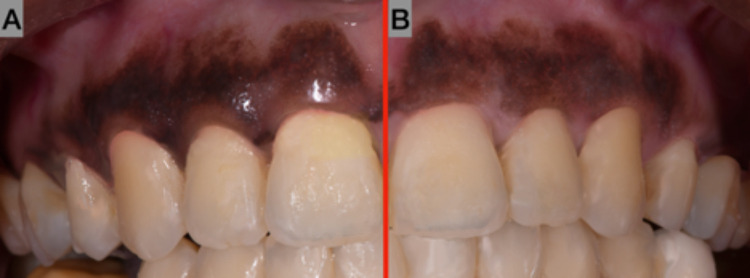
目的:比较极化和非极化口腔内摄影测量牙龈黑色素沉着的效度和信度。材料与方法:对10例牙龈色素沉着患者进行病例系列研究。收集了976张偏光和非偏光图像样本,在牙龈边缘以上两排进行分析。这些图像是在脱色手术之前和一年后拍摄的。三位独立的评估者评估了这些照片(一位正畸医生、一位普通牙医和一位门外汉)。采用Dummett Oral Pigmentation Index (DOPI)和Gingival Melanosis Record (GMR)指数检测牙龈色素沉着水平。结果:研究发现,在脱色前后拍摄的偏振和非偏振图像无显著差异。两种成像方法从评估者那里得到了相似的分数。正畸医生比外行人和普通牙医识别出更多的色素玻片。结论:偏光法和非偏光法均可用于评估牙龈色素沉着。然而,需要进一步的研究来证实这一发现并检查其他因素。
Validity and Reliability of Polarized vs Non-Polarized Digital Images for Measuring Gingival Melanin Pigmentation.
Aim: This study aimed to compare the validity and reliability of polarized and non-polarized intraoral photography for the measurement of gingival melanin pigmentation.
Materials and methods: A case series study was conducted on ten patients scheduled for gingival depigmentation. A total of 976 polarized and non-polarized image samples were collected, capturing two rows above the gingival margin, for analysis. These images were taken both before and one year after the depigmentation procedure. Three independent evaluators assessed the photographs (an orthodontist, a general dentist, and a layperson). The Dummett Oral Pigmentation Index (DOPI) and Gingival Melanosis Record (GMR) indices were used to measure the level of gingival pigmentation.
Results: The study found no significant differences between polarized and non-polarized images taken before and after depigmentation. Both methods of imaging received similar scores from the evaluators. The orthodontist identified more pigmented slides than the layperson and the general dentist.
Conclusion: Both polarized and non-polarized photographic methods may be used for assessing gingival pigmentation. However, further research is warranted to confirm this finding and examine additional factors.

 求助内容:
求助内容: 应助结果提醒方式:


