旁矢状面囊性病变可能起源于皮层静脉壁周围的脑膜鞘。
IF 3.2
3区 医学
Q2 RADIOLOGY, NUCLEAR MEDICINE & MEDICAL IMAGING
引用次数: 1
摘要
据报道,在静脉注射后4小时,矢状旁硬脑膜附近的静脉周围囊性结构与钆基造影剂的渗漏有关。这种囊性结构的起源尚不清楚。在阅读许多病例的磁共振脑池造影时,我们注意到一些囊性结构似乎连接到静脉周围的腹膜下间隙。这一新的影像学发现可能有助于未来中枢神经系统废物清除系统的研究。本文章由计算机程序翻译,如有差异,请以英文原文为准。
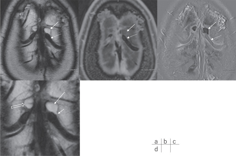
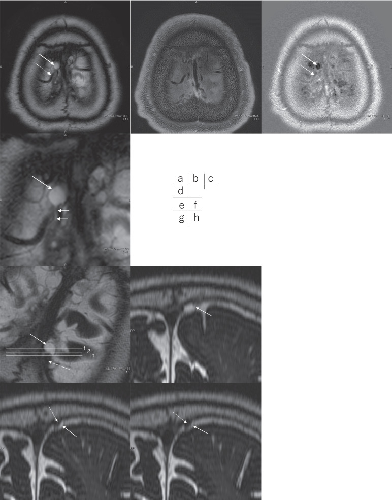
Parasagittal Cystic Lesions May Arise from the Pial Sheath around the Cortical Venous Wall.
It has been reported that perivenous cystic structures near the parasagittal dura are associated with the leakage of gadolinium-based contrast agents at 4 hours after intravenous administration. The origin of such cystic structures remains unknown. While reading many cases of MR cisternography, we noticed that some of the cystic structures appeared to connect to the perivenous subpial space. This new imaging finding might facilitate future research of the waste clearance system for the central nervous system.
求助全文
通过发布文献求助,成功后即可免费获取论文全文。
去求助
来源期刊
Magnetic Resonance in Medical Sciences
RADIOLOGY, NUCLEAR MEDICINE & MEDICAL IMAGING-
CiteScore
5.80
自引率
20.00%
发文量
71
审稿时长
>12 weeks
期刊介绍:
Magnetic Resonance in Medical Sciences (MRMS or Magn
Reson Med Sci) is an international journal pursuing the
publication of original articles contributing to the progress
of magnetic resonance in the field of biomedical sciences
including technical developments and clinical applications.
MRMS is an official journal of the Japanese Society for
Magnetic Resonance in Medicine (JSMRM).
 求助内容:
求助内容: 应助结果提醒方式:


