{"title":"组织中SARS-CoV-2检测方法。","authors":"Saskia von Stillfried, Peter Boor","doi":"10.1007/s00292-021-00920-1","DOIUrl":null,"url":null,"abstract":"<p><strong>Background: </strong>Analyses for the presence of SARS-CoV‑2 in the tissues of COVID-19 patients is important in order to improve our understanding of the disease pathophysiology for interpretation of diagnostic histopathological findings in autopsies, biopsies, or surgical specimens and to assess the potential for occupational infectious hazard.</p><p><strong>Material and methods: </strong>In this review we identified 136 published studies in PubMed's curated literature database LitCovid on SARS-CoV‑2 detection methods in tissues and evaluated them regarding sources of error, specificity, and sensitivity of the methods, taking into account our own experience.</p><p><strong>Results: </strong>Currently, no sufficiently specific histomorphological alterations or diagnostic features for COVID-19 are known. Therefore, three approaches for SARS-CoV‑2 detection are used: RNA, proteins/antigens, or morphological detection by electron microscopy. In the preanalytical phase, the dominant source of error is tissue quality, especially the different intervals between sample collection and processing or fixation (and its duration) and specifically the interval between death and sample collection in autopsies. However, this information is found in less than half of the studies (e.g., in only 42% of autopsy studies). Our own experience and first studies prove the significantly higher sensitivity and specificity of RNA-based detection methods compared to antigen or protein detection by immunohistochemistry or immunofluorescence. Detection by electron microscopy is time consuming and difficult to interpret.</p><p><strong>Conclusions: </strong>Different methods are available for the detection of SARS-CoV‑2 in tissue. Currently, RNA detection by RT-PCR is the method of choice. However, extensive validation studies and method harmonization are not available and are absolutely necessary.</p>","PeriodicalId":54641,"journal":{"name":"Pathologe","volume":"42 Suppl 1","pages":"81-88"},"PeriodicalIF":0.0000,"publicationDate":"2021-11-01","publicationTypes":"Journal Article","fieldsOfStudy":null,"isOpenAccess":false,"openAccessPdf":"https://sci-hub-pdf.com/10.1007/s00292-021-00920-1","citationCount":"11","resultStr":"{\"title\":\"Detection methods for SARS-CoV-2 in tissue.\",\"authors\":\"Saskia von Stillfried, Peter Boor\",\"doi\":\"10.1007/s00292-021-00920-1\",\"DOIUrl\":null,\"url\":null,\"abstract\":\"<p><strong>Background: </strong>Analyses for the presence of SARS-CoV‑2 in the tissues of COVID-19 patients is important in order to improve our understanding of the disease pathophysiology for interpretation of diagnostic histopathological findings in autopsies, biopsies, or surgical specimens and to assess the potential for occupational infectious hazard.</p><p><strong>Material and methods: </strong>In this review we identified 136 published studies in PubMed's curated literature database LitCovid on SARS-CoV‑2 detection methods in tissues and evaluated them regarding sources of error, specificity, and sensitivity of the methods, taking into account our own experience.</p><p><strong>Results: </strong>Currently, no sufficiently specific histomorphological alterations or diagnostic features for COVID-19 are known. Therefore, three approaches for SARS-CoV‑2 detection are used: RNA, proteins/antigens, or morphological detection by electron microscopy. In the preanalytical phase, the dominant source of error is tissue quality, especially the different intervals between sample collection and processing or fixation (and its duration) and specifically the interval between death and sample collection in autopsies. However, this information is found in less than half of the studies (e.g., in only 42% of autopsy studies). Our own experience and first studies prove the significantly higher sensitivity and specificity of RNA-based detection methods compared to antigen or protein detection by immunohistochemistry or immunofluorescence. Detection by electron microscopy is time consuming and difficult to interpret.</p><p><strong>Conclusions: </strong>Different methods are available for the detection of SARS-CoV‑2 in tissue. Currently, RNA detection by RT-PCR is the method of choice. However, extensive validation studies and method harmonization are not available and are absolutely necessary.</p>\",\"PeriodicalId\":54641,\"journal\":{\"name\":\"Pathologe\",\"volume\":\"42 Suppl 1\",\"pages\":\"81-88\"},\"PeriodicalIF\":0.0000,\"publicationDate\":\"2021-11-01\",\"publicationTypes\":\"Journal Article\",\"fieldsOfStudy\":null,\"isOpenAccess\":false,\"openAccessPdf\":\"https://sci-hub-pdf.com/10.1007/s00292-021-00920-1\",\"citationCount\":\"11\",\"resultStr\":null,\"platform\":\"Semanticscholar\",\"paperid\":null,\"PeriodicalName\":\"Pathologe\",\"FirstCategoryId\":\"3\",\"ListUrlMain\":\"https://doi.org/10.1007/s00292-021-00920-1\",\"RegionNum\":4,\"RegionCategory\":\"医学\",\"ArticlePicture\":[],\"TitleCN\":null,\"AbstractTextCN\":null,\"PMCID\":null,\"EPubDate\":\"\",\"PubModel\":\"\",\"JCR\":\"Q3\",\"JCRName\":\"Medicine\",\"Score\":null,\"Total\":0}","platform":"Semanticscholar","paperid":null,"PeriodicalName":"Pathologe","FirstCategoryId":"3","ListUrlMain":"https://doi.org/10.1007/s00292-021-00920-1","RegionNum":4,"RegionCategory":"医学","ArticlePicture":[],"TitleCN":null,"AbstractTextCN":null,"PMCID":null,"EPubDate":"","PubModel":"","JCR":"Q3","JCRName":"Medicine","Score":null,"Total":0}
Background: Analyses for the presence of SARS-CoV‑2 in the tissues of COVID-19 patients is important in order to improve our understanding of the disease pathophysiology for interpretation of diagnostic histopathological findings in autopsies, biopsies, or surgical specimens and to assess the potential for occupational infectious hazard.
Material and methods: In this review we identified 136 published studies in PubMed's curated literature database LitCovid on SARS-CoV‑2 detection methods in tissues and evaluated them regarding sources of error, specificity, and sensitivity of the methods, taking into account our own experience.
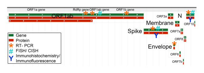
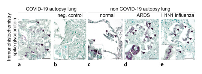
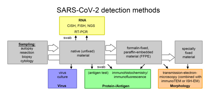
Results: Currently, no sufficiently specific histomorphological alterations or diagnostic features for COVID-19 are known. Therefore, three approaches for SARS-CoV‑2 detection are used: RNA, proteins/antigens, or morphological detection by electron microscopy. In the preanalytical phase, the dominant source of error is tissue quality, especially the different intervals between sample collection and processing or fixation (and its duration) and specifically the interval between death and sample collection in autopsies. However, this information is found in less than half of the studies (e.g., in only 42% of autopsy studies). Our own experience and first studies prove the significantly higher sensitivity and specificity of RNA-based detection methods compared to antigen or protein detection by immunohistochemistry or immunofluorescence. Detection by electron microscopy is time consuming and difficult to interpret.
Conclusions: Different methods are available for the detection of SARS-CoV‑2 in tissue. Currently, RNA detection by RT-PCR is the method of choice. However, extensive validation studies and method harmonization are not available and are absolutely necessary.
期刊介绍:
Der Pathologe is an internationally recognized journal and combines practical relevance with scientific competence. The journal informs all pathologists working on departments and institutes as well as morphologically interested scientists about developments in the field of pathology.
The journal serves both the scientific exchange and the continuing education of pathologists.
Comprehensive reviews on a specific topical issue focus on providing evidenced based information under consideration of practical experience.
Freely submitted original papers allow the presentation of important clinical studies and serve the scientific exchange.
 求助内容:
求助内容: 应助结果提醒方式:


