Dhananjay Yellajoshyula, Sunday Opeyemi, William T Dauer, Samuel S Pappas
{"title":"张力障碍小鼠模型纹状体突触异常成熟和分泌通路改变的遗传证据。","authors":"Dhananjay Yellajoshyula, Sunday Opeyemi, William T Dauer, Samuel S Pappas","doi":"10.3389/dyst.2022.10892","DOIUrl":null,"url":null,"abstract":"<p><p>Animal models of DYT-TOR1A dystonia consistently demonstrate abnormalities of striatal cholinergic function, but the molecular pathways underlying this pathophysiology are unclear. To probe these molecular pathways in a genetic model of DYT-TOR1A, we performed laser microdissection in juvenile mice to isolate striatal cholinergic interneurons and non-cholinergic striatal tissue largely comprising spiny projection neurons during maturation. Both cholinergic and GABAergic enriched samples demonstrated a defined set of gene expression changes consistent with a role of torsinA in the secretory pathway. GABAergic enriched striatum samples also showed alteration to genes regulating synaptic transmission and an upregulation of activity dependent immediate early genes. Reconstruction of Golgi-Cox stained striatal spiny projection neurons from adult mice demonstrated significantly increased spiny density, suggesting that torsinA null striatal neurons have increased excitability during striatal maturation and long lasting increases in afferent input. These findings are consistent with a developmental role for torsinA in the secretory pathway and link torsinA loss of function with functional and structural changes of striatal cholinergic and GABAergic neurons. These transcriptomic datasets are freely available as a resource for future studies of torsinA loss of function-mediated striatal dysfunction.</p>","PeriodicalId":72853,"journal":{"name":"Dystonia","volume":"1 ","pages":""},"PeriodicalIF":0.0000,"publicationDate":"2022-01-01","publicationTypes":"Journal Article","fieldsOfStudy":null,"isOpenAccess":false,"openAccessPdf":"https://www.ncbi.nlm.nih.gov/pmc/articles/PMC9980434/pdf/","citationCount":"1","resultStr":"{\"title\":\"Genetic evidence of aberrant striatal synaptic maturation and secretory pathway alteration in a dystonia mouse model.\",\"authors\":\"Dhananjay Yellajoshyula, Sunday Opeyemi, William T Dauer, Samuel S Pappas\",\"doi\":\"10.3389/dyst.2022.10892\",\"DOIUrl\":null,\"url\":null,\"abstract\":\"<p><p>Animal models of DYT-TOR1A dystonia consistently demonstrate abnormalities of striatal cholinergic function, but the molecular pathways underlying this pathophysiology are unclear. To probe these molecular pathways in a genetic model of DYT-TOR1A, we performed laser microdissection in juvenile mice to isolate striatal cholinergic interneurons and non-cholinergic striatal tissue largely comprising spiny projection neurons during maturation. Both cholinergic and GABAergic enriched samples demonstrated a defined set of gene expression changes consistent with a role of torsinA in the secretory pathway. GABAergic enriched striatum samples also showed alteration to genes regulating synaptic transmission and an upregulation of activity dependent immediate early genes. Reconstruction of Golgi-Cox stained striatal spiny projection neurons from adult mice demonstrated significantly increased spiny density, suggesting that torsinA null striatal neurons have increased excitability during striatal maturation and long lasting increases in afferent input. These findings are consistent with a developmental role for torsinA in the secretory pathway and link torsinA loss of function with functional and structural changes of striatal cholinergic and GABAergic neurons. These transcriptomic datasets are freely available as a resource for future studies of torsinA loss of function-mediated striatal dysfunction.</p>\",\"PeriodicalId\":72853,\"journal\":{\"name\":\"Dystonia\",\"volume\":\"1 \",\"pages\":\"\"},\"PeriodicalIF\":0.0000,\"publicationDate\":\"2022-01-01\",\"publicationTypes\":\"Journal Article\",\"fieldsOfStudy\":null,\"isOpenAccess\":false,\"openAccessPdf\":\"https://www.ncbi.nlm.nih.gov/pmc/articles/PMC9980434/pdf/\",\"citationCount\":\"1\",\"resultStr\":null,\"platform\":\"Semanticscholar\",\"paperid\":null,\"PeriodicalName\":\"Dystonia\",\"FirstCategoryId\":\"1085\",\"ListUrlMain\":\"https://doi.org/10.3389/dyst.2022.10892\",\"RegionNum\":0,\"RegionCategory\":null,\"ArticlePicture\":[],\"TitleCN\":null,\"AbstractTextCN\":null,\"PMCID\":null,\"EPubDate\":\"\",\"PubModel\":\"\",\"JCR\":\"\",\"JCRName\":\"\",\"Score\":null,\"Total\":0}","platform":"Semanticscholar","paperid":null,"PeriodicalName":"Dystonia","FirstCategoryId":"1085","ListUrlMain":"https://doi.org/10.3389/dyst.2022.10892","RegionNum":0,"RegionCategory":null,"ArticlePicture":[],"TitleCN":null,"AbstractTextCN":null,"PMCID":null,"EPubDate":"","PubModel":"","JCR":"","JCRName":"","Score":null,"Total":0}
Genetic evidence of aberrant striatal synaptic maturation and secretory pathway alteration in a dystonia mouse model.
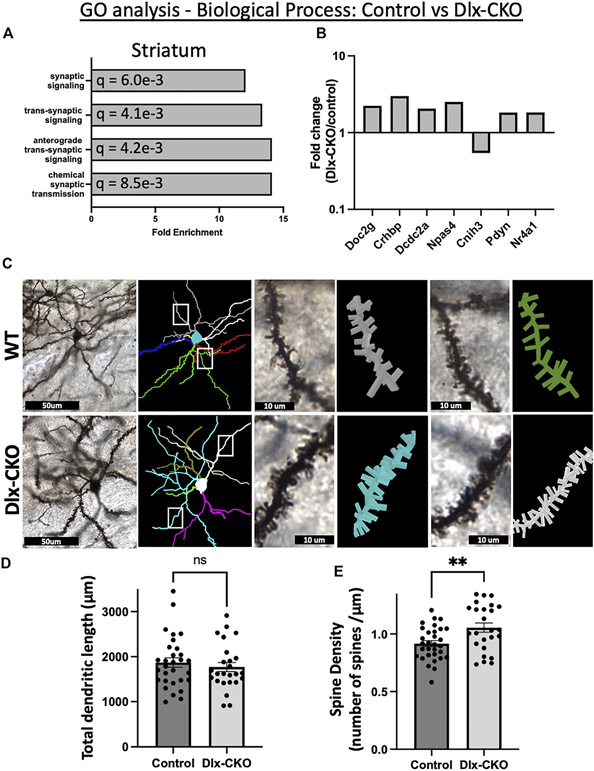
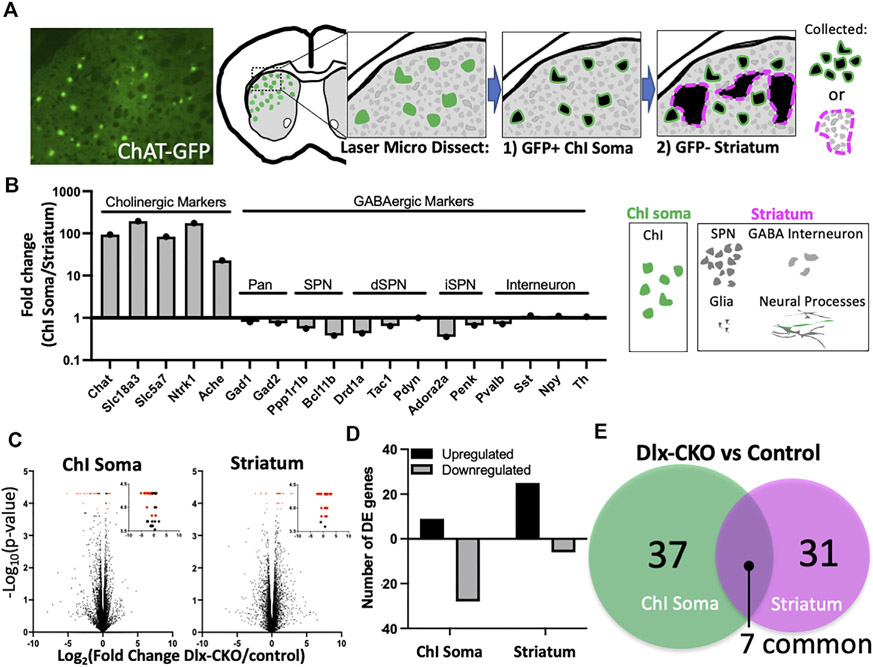
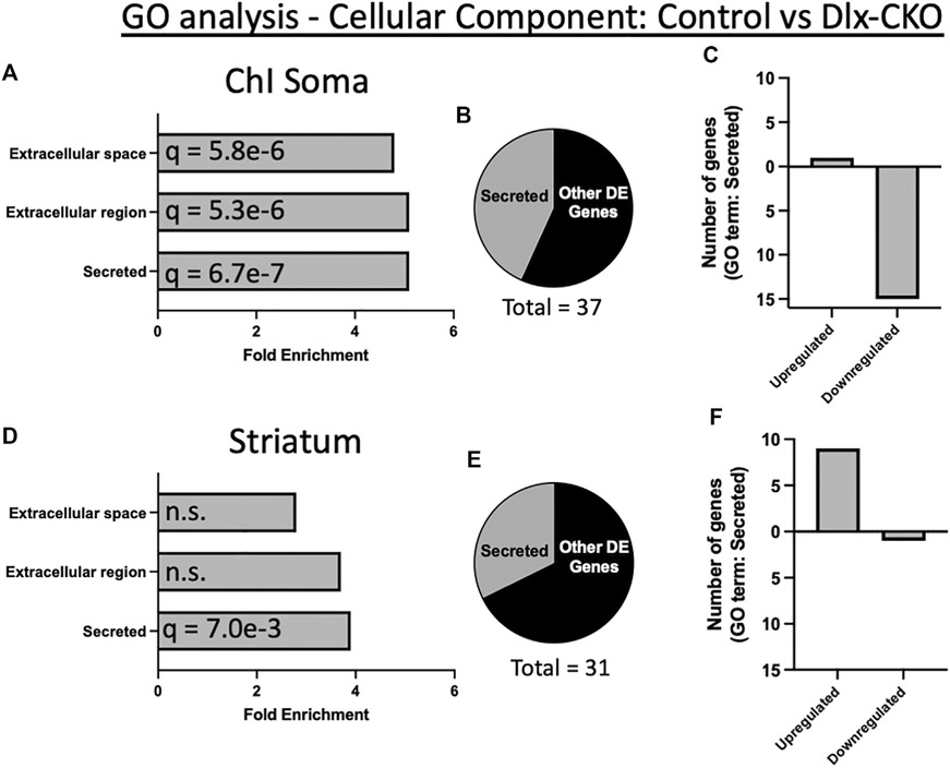
Animal models of DYT-TOR1A dystonia consistently demonstrate abnormalities of striatal cholinergic function, but the molecular pathways underlying this pathophysiology are unclear. To probe these molecular pathways in a genetic model of DYT-TOR1A, we performed laser microdissection in juvenile mice to isolate striatal cholinergic interneurons and non-cholinergic striatal tissue largely comprising spiny projection neurons during maturation. Both cholinergic and GABAergic enriched samples demonstrated a defined set of gene expression changes consistent with a role of torsinA in the secretory pathway. GABAergic enriched striatum samples also showed alteration to genes regulating synaptic transmission and an upregulation of activity dependent immediate early genes. Reconstruction of Golgi-Cox stained striatal spiny projection neurons from adult mice demonstrated significantly increased spiny density, suggesting that torsinA null striatal neurons have increased excitability during striatal maturation and long lasting increases in afferent input. These findings are consistent with a developmental role for torsinA in the secretory pathway and link torsinA loss of function with functional and structural changes of striatal cholinergic and GABAergic neurons. These transcriptomic datasets are freely available as a resource for future studies of torsinA loss of function-mediated striatal dysfunction.

 求助内容:
求助内容: 应助结果提醒方式:


