Laura Calderan, Flavia Carton, Ilaria Andreana, Valeria Bincoletto, Silvia Arpicco, Barbara Stella, Manuela Malatesta
{"title":"追踪骨骼肌内荧光纳米颗粒的离体实验系统。","authors":"Laura Calderan, Flavia Carton, Ilaria Andreana, Valeria Bincoletto, Silvia Arpicco, Barbara Stella, Manuela Malatesta","doi":"10.4081/ejh.2023.3596","DOIUrl":null,"url":null,"abstract":"<p><p>The development of novel nanoconstructs for biomedical applications requires the assessment of their biodistribution, metabolism and clearance in single cells, organs and entire organisms in a living environment. To reduce the number of in vivo experiments performed and to refine the methods used, in accordance with the 3Rs principle, this work proposes an ex vivo experimental system to monitor, using fluorescence microscopy, the distribution of nanoparticles in explanted murine skeletal muscle maintained in a bioreactor that can preserve the structural and functional features of the organ for long periods of time. Fluorescently-labelled liposomes and poly(lactide-co-glycolide) (PLGA)-based nanoparticles were injected into the intact soleus muscle (in the distal region close to the tendon) immediately after explants, and their distribution was analysed at increasing incubation times in cross cryosections from the proximal region of the belly. Both nanocarriers were clearly recognized in the muscle and were found to enter and migrate inside the myofibres, whereas their migration in the connective tissue seemed to be limited. In addition, some fluorescent signals were observed inside the macrophages, demonstrating the physiological clearance of the nanocarriers that did not enter the myofibres. Our ex vivo system therefore provides more information than previous in vitro experiments on cultured muscle cells, highlighting the need for the appropriate functionalization of nanocarriers if myofibre targeting is to be improved.</p>","PeriodicalId":50487,"journal":{"name":"European Journal of Histochemistry","volume":"67 1","pages":""},"PeriodicalIF":2.1000,"publicationDate":"2023-01-02","publicationTypes":"Journal Article","fieldsOfStudy":null,"isOpenAccess":false,"openAccessPdf":"https://ftp.ncbi.nlm.nih.gov/pub/pmc/oa_pdf/20/a6/ejh-67-1-3596.PMC9827424.pdf","citationCount":"1","resultStr":"{\"title\":\"An ex vivo experimental system to track fluorescent nanoparticles inside skeletal muscle.\",\"authors\":\"Laura Calderan, Flavia Carton, Ilaria Andreana, Valeria Bincoletto, Silvia Arpicco, Barbara Stella, Manuela Malatesta\",\"doi\":\"10.4081/ejh.2023.3596\",\"DOIUrl\":null,\"url\":null,\"abstract\":\"<p><p>The development of novel nanoconstructs for biomedical applications requires the assessment of their biodistribution, metabolism and clearance in single cells, organs and entire organisms in a living environment. To reduce the number of in vivo experiments performed and to refine the methods used, in accordance with the 3Rs principle, this work proposes an ex vivo experimental system to monitor, using fluorescence microscopy, the distribution of nanoparticles in explanted murine skeletal muscle maintained in a bioreactor that can preserve the structural and functional features of the organ for long periods of time. Fluorescently-labelled liposomes and poly(lactide-co-glycolide) (PLGA)-based nanoparticles were injected into the intact soleus muscle (in the distal region close to the tendon) immediately after explants, and their distribution was analysed at increasing incubation times in cross cryosections from the proximal region of the belly. Both nanocarriers were clearly recognized in the muscle and were found to enter and migrate inside the myofibres, whereas their migration in the connective tissue seemed to be limited. In addition, some fluorescent signals were observed inside the macrophages, demonstrating the physiological clearance of the nanocarriers that did not enter the myofibres. Our ex vivo system therefore provides more information than previous in vitro experiments on cultured muscle cells, highlighting the need for the appropriate functionalization of nanocarriers if myofibre targeting is to be improved.</p>\",\"PeriodicalId\":50487,\"journal\":{\"name\":\"European Journal of Histochemistry\",\"volume\":\"67 1\",\"pages\":\"\"},\"PeriodicalIF\":2.1000,\"publicationDate\":\"2023-01-02\",\"publicationTypes\":\"Journal Article\",\"fieldsOfStudy\":null,\"isOpenAccess\":false,\"openAccessPdf\":\"https://ftp.ncbi.nlm.nih.gov/pub/pmc/oa_pdf/20/a6/ejh-67-1-3596.PMC9827424.pdf\",\"citationCount\":\"1\",\"resultStr\":null,\"platform\":\"Semanticscholar\",\"paperid\":null,\"PeriodicalName\":\"European Journal of Histochemistry\",\"FirstCategoryId\":\"99\",\"ListUrlMain\":\"https://doi.org/10.4081/ejh.2023.3596\",\"RegionNum\":4,\"RegionCategory\":\"生物学\",\"ArticlePicture\":[],\"TitleCN\":null,\"AbstractTextCN\":null,\"PMCID\":null,\"EPubDate\":\"\",\"PubModel\":\"\",\"JCR\":\"Q4\",\"JCRName\":\"CELL BIOLOGY\",\"Score\":null,\"Total\":0}","platform":"Semanticscholar","paperid":null,"PeriodicalName":"European Journal of Histochemistry","FirstCategoryId":"99","ListUrlMain":"https://doi.org/10.4081/ejh.2023.3596","RegionNum":4,"RegionCategory":"生物学","ArticlePicture":[],"TitleCN":null,"AbstractTextCN":null,"PMCID":null,"EPubDate":"","PubModel":"","JCR":"Q4","JCRName":"CELL BIOLOGY","Score":null,"Total":0}
An ex vivo experimental system to track fluorescent nanoparticles inside skeletal muscle.
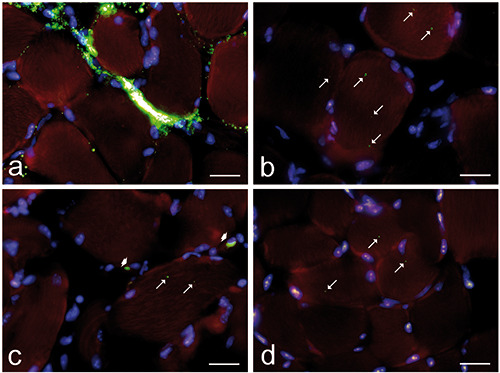
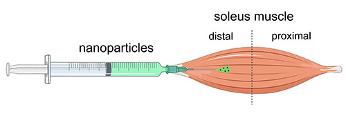
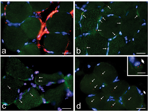
The development of novel nanoconstructs for biomedical applications requires the assessment of their biodistribution, metabolism and clearance in single cells, organs and entire organisms in a living environment. To reduce the number of in vivo experiments performed and to refine the methods used, in accordance with the 3Rs principle, this work proposes an ex vivo experimental system to monitor, using fluorescence microscopy, the distribution of nanoparticles in explanted murine skeletal muscle maintained in a bioreactor that can preserve the structural and functional features of the organ for long periods of time. Fluorescently-labelled liposomes and poly(lactide-co-glycolide) (PLGA)-based nanoparticles were injected into the intact soleus muscle (in the distal region close to the tendon) immediately after explants, and their distribution was analysed at increasing incubation times in cross cryosections from the proximal region of the belly. Both nanocarriers were clearly recognized in the muscle and were found to enter and migrate inside the myofibres, whereas their migration in the connective tissue seemed to be limited. In addition, some fluorescent signals were observed inside the macrophages, demonstrating the physiological clearance of the nanocarriers that did not enter the myofibres. Our ex vivo system therefore provides more information than previous in vitro experiments on cultured muscle cells, highlighting the need for the appropriate functionalization of nanocarriers if myofibre targeting is to be improved.
期刊介绍:
The Journal publishes original papers concerning investigations by histochemical and immunohistochemical methods, and performed with the aid of light, super-resolution and electron microscopy, cytometry and imaging techniques. Coverage extends to:
functional cell and tissue biology in animals and plants;
cell differentiation and death;
cell-cell interaction and molecular trafficking;
biology of cell development and senescence;
nerve and muscle cell biology;
cellular basis of diseases.
The histochemical approach is nowadays essentially aimed at locating molecules in the very place where they exert their biological roles, and at describing dynamically specific chemical activities in living cells. Basic research on cell functional organization is essential for understanding the mechanisms underlying major biological processes such as differentiation, the control of tissue homeostasis, and the regulation of normal and tumor cell growth. Even more than in the past, the European Journal of Histochemistry, as a journal of functional cytology, represents the venue where cell scientists may present and discuss their original results, technical improvements and theories.
 求助内容:
求助内容: 应助结果提醒方式:


