Grith Højfeldt, Trent Sorenson, Alana Gonzales, Michael Kjaer, Jesper L Andersen, Abigail L Mackey
{"title":"肌纤维分支的融合是健康人体骨骼肌再生的生理特征。","authors":"Grith Højfeldt, Trent Sorenson, Alana Gonzales, Michael Kjaer, Jesper L Andersen, Abigail L Mackey","doi":"10.1186/s13395-023-00322-2","DOIUrl":null,"url":null,"abstract":"<p><strong>Background: </strong>The occurrence of hyperplasia, through myofibre splitting, remains a widely debated phenomenon. Structural alterations and fibre typing of skeletal muscle fibres, as seen during regeneration and in certain muscle diseases, can be challenging to interpret. Neuromuscular electrical stimulation can induce myofibre necrosis followed by changes in spatial and temporal cellular processes. Thirty days following electrical stimulation, remnants of regeneration can be seen in the myofibre and its basement membrane as the presence of small myofibres and encroachment of sarcolemma and basement membrane (suggestive of myofibre branching/splitting). The purpose of this study was to investigate myofibre branching and fibre type in a systematic manner in human skeletal muscle undergoing adult regenerative myogenesis.</p><p><strong>Methods: </strong>Electrical stimulation was used to induce myofibre necrosis to the vastus lateralis muscle of one leg in 5 young healthy males. Muscle tissue samples were collected from the stimulated leg 30 days later and from the control leg for comparison. Biopsies were sectioned and stained for dystrophin and laminin to label the sarcolemma and basement membrane, respectively, as well as ATPase, and antibodies against types I and II myosin, and embryonic and neonatal myosin. Myofibre branches were followed through 22 serial Sects. (264 μm). Single fibres and tissue blocks were examined by confocal and electron microscopy, respectively.</p><p><strong>Results: </strong>Regular branching of small myofibre segments was observed (median length 144 μm), most of which were observed to fuse further along the parent fibre. Central nuclei were frequently observed at the point of branching/fusion. The branch commonly presented with a more immature profile (nestin + , neonatal myosin + , disorganised myofilaments) than the parent myofibre, together suggesting fusion of the branch, rather than splitting. Of the 210 regenerating muscle fibres evaluated, 99.5% were type II fibres, indicating preferential damage to type II fibres with our protocol. Furthermore, these fibres demonstrated 7 different stages of \"fibre-type\" profiles.</p><p><strong>Conclusions: </strong>By studying the regenerating tissue 30 days later with a range of microscopy techniques, we find that so-called myofibre branching or splitting is more likely to be fusion of myotubes and is therefore explained by incomplete regeneration after a necrosis-inducing event.</p>","PeriodicalId":21747,"journal":{"name":"Skeletal Muscle","volume":"13 1","pages":"13"},"PeriodicalIF":4.4000,"publicationDate":"2023-08-12","publicationTypes":"Journal Article","fieldsOfStudy":null,"isOpenAccess":false,"openAccessPdf":"https://www.ncbi.nlm.nih.gov/pmc/articles/PMC10422711/pdf/","citationCount":"0","resultStr":"{\"title\":\"Fusion of myofibre branches is a physiological feature of healthy human skeletal muscle regeneration.\",\"authors\":\"Grith Højfeldt, Trent Sorenson, Alana Gonzales, Michael Kjaer, Jesper L Andersen, Abigail L Mackey\",\"doi\":\"10.1186/s13395-023-00322-2\",\"DOIUrl\":null,\"url\":null,\"abstract\":\"<p><strong>Background: </strong>The occurrence of hyperplasia, through myofibre splitting, remains a widely debated phenomenon. Structural alterations and fibre typing of skeletal muscle fibres, as seen during regeneration and in certain muscle diseases, can be challenging to interpret. Neuromuscular electrical stimulation can induce myofibre necrosis followed by changes in spatial and temporal cellular processes. Thirty days following electrical stimulation, remnants of regeneration can be seen in the myofibre and its basement membrane as the presence of small myofibres and encroachment of sarcolemma and basement membrane (suggestive of myofibre branching/splitting). The purpose of this study was to investigate myofibre branching and fibre type in a systematic manner in human skeletal muscle undergoing adult regenerative myogenesis.</p><p><strong>Methods: </strong>Electrical stimulation was used to induce myofibre necrosis to the vastus lateralis muscle of one leg in 5 young healthy males. Muscle tissue samples were collected from the stimulated leg 30 days later and from the control leg for comparison. Biopsies were sectioned and stained for dystrophin and laminin to label the sarcolemma and basement membrane, respectively, as well as ATPase, and antibodies against types I and II myosin, and embryonic and neonatal myosin. Myofibre branches were followed through 22 serial Sects. (264 μm). Single fibres and tissue blocks were examined by confocal and electron microscopy, respectively.</p><p><strong>Results: </strong>Regular branching of small myofibre segments was observed (median length 144 μm), most of which were observed to fuse further along the parent fibre. Central nuclei were frequently observed at the point of branching/fusion. The branch commonly presented with a more immature profile (nestin + , neonatal myosin + , disorganised myofilaments) than the parent myofibre, together suggesting fusion of the branch, rather than splitting. Of the 210 regenerating muscle fibres evaluated, 99.5% were type II fibres, indicating preferential damage to type II fibres with our protocol. Furthermore, these fibres demonstrated 7 different stages of \\\"fibre-type\\\" profiles.</p><p><strong>Conclusions: </strong>By studying the regenerating tissue 30 days later with a range of microscopy techniques, we find that so-called myofibre branching or splitting is more likely to be fusion of myotubes and is therefore explained by incomplete regeneration after a necrosis-inducing event.</p>\",\"PeriodicalId\":21747,\"journal\":{\"name\":\"Skeletal Muscle\",\"volume\":\"13 1\",\"pages\":\"13\"},\"PeriodicalIF\":4.4000,\"publicationDate\":\"2023-08-12\",\"publicationTypes\":\"Journal Article\",\"fieldsOfStudy\":null,\"isOpenAccess\":false,\"openAccessPdf\":\"https://www.ncbi.nlm.nih.gov/pmc/articles/PMC10422711/pdf/\",\"citationCount\":\"0\",\"resultStr\":null,\"platform\":\"Semanticscholar\",\"paperid\":null,\"PeriodicalName\":\"Skeletal Muscle\",\"FirstCategoryId\":\"3\",\"ListUrlMain\":\"https://doi.org/10.1186/s13395-023-00322-2\",\"RegionNum\":2,\"RegionCategory\":\"医学\",\"ArticlePicture\":[],\"TitleCN\":null,\"AbstractTextCN\":null,\"PMCID\":null,\"EPubDate\":\"\",\"PubModel\":\"\",\"JCR\":\"Q2\",\"JCRName\":\"CELL BIOLOGY\",\"Score\":null,\"Total\":0}","platform":"Semanticscholar","paperid":null,"PeriodicalName":"Skeletal Muscle","FirstCategoryId":"3","ListUrlMain":"https://doi.org/10.1186/s13395-023-00322-2","RegionNum":2,"RegionCategory":"医学","ArticlePicture":[],"TitleCN":null,"AbstractTextCN":null,"PMCID":null,"EPubDate":"","PubModel":"","JCR":"Q2","JCRName":"CELL BIOLOGY","Score":null,"Total":0}
Fusion of myofibre branches is a physiological feature of healthy human skeletal muscle regeneration.
Background: The occurrence of hyperplasia, through myofibre splitting, remains a widely debated phenomenon. Structural alterations and fibre typing of skeletal muscle fibres, as seen during regeneration and in certain muscle diseases, can be challenging to interpret. Neuromuscular electrical stimulation can induce myofibre necrosis followed by changes in spatial and temporal cellular processes. Thirty days following electrical stimulation, remnants of regeneration can be seen in the myofibre and its basement membrane as the presence of small myofibres and encroachment of sarcolemma and basement membrane (suggestive of myofibre branching/splitting). The purpose of this study was to investigate myofibre branching and fibre type in a systematic manner in human skeletal muscle undergoing adult regenerative myogenesis.
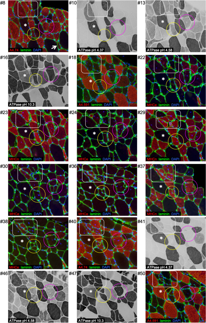
Methods: Electrical stimulation was used to induce myofibre necrosis to the vastus lateralis muscle of one leg in 5 young healthy males. Muscle tissue samples were collected from the stimulated leg 30 days later and from the control leg for comparison. Biopsies were sectioned and stained for dystrophin and laminin to label the sarcolemma and basement membrane, respectively, as well as ATPase, and antibodies against types I and II myosin, and embryonic and neonatal myosin. Myofibre branches were followed through 22 serial Sects. (264 μm). Single fibres and tissue blocks were examined by confocal and electron microscopy, respectively.
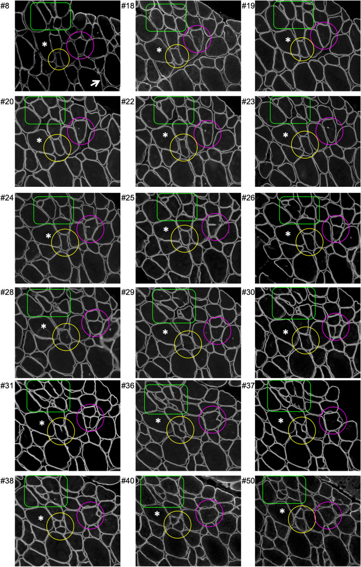
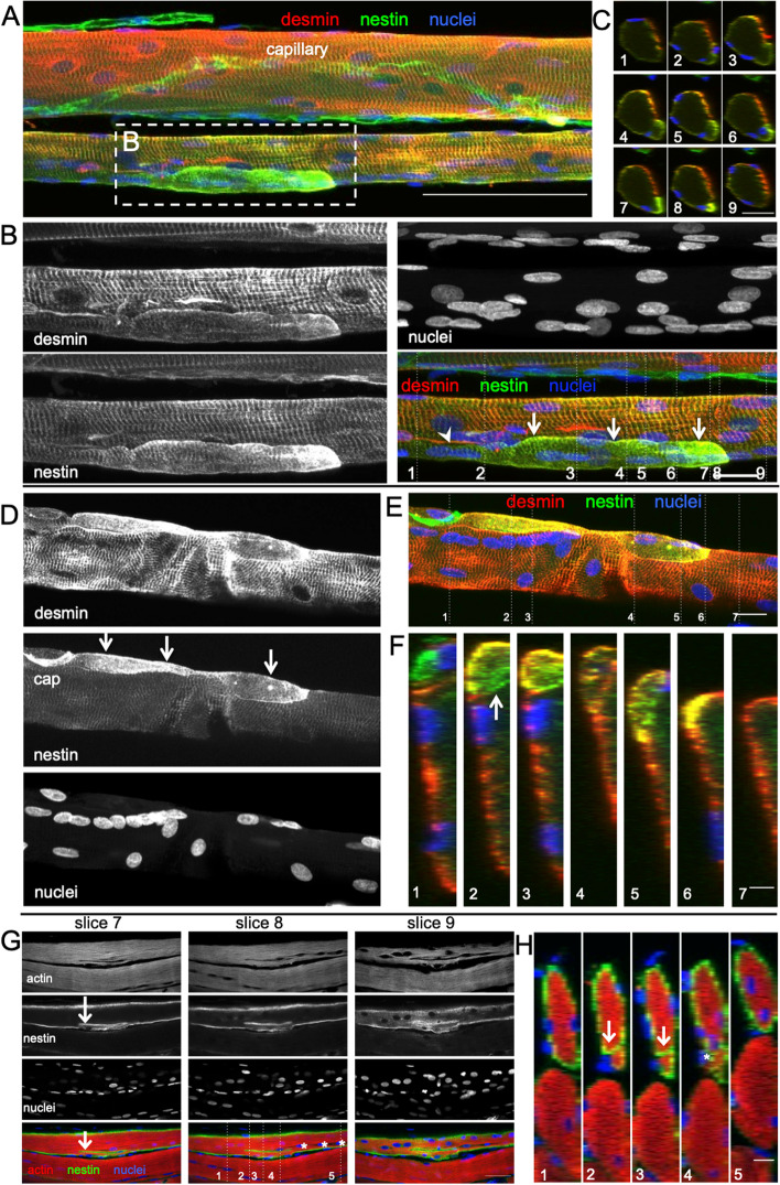
Results: Regular branching of small myofibre segments was observed (median length 144 μm), most of which were observed to fuse further along the parent fibre. Central nuclei were frequently observed at the point of branching/fusion. The branch commonly presented with a more immature profile (nestin + , neonatal myosin + , disorganised myofilaments) than the parent myofibre, together suggesting fusion of the branch, rather than splitting. Of the 210 regenerating muscle fibres evaluated, 99.5% were type II fibres, indicating preferential damage to type II fibres with our protocol. Furthermore, these fibres demonstrated 7 different stages of "fibre-type" profiles.
Conclusions: By studying the regenerating tissue 30 days later with a range of microscopy techniques, we find that so-called myofibre branching or splitting is more likely to be fusion of myotubes and is therefore explained by incomplete regeneration after a necrosis-inducing event.
期刊介绍:
The only open access journal in its field, Skeletal Muscle publishes novel, cutting-edge research and technological advancements that investigate the molecular mechanisms underlying the biology of skeletal muscle. Reflecting the breadth of research in this area, the journal welcomes manuscripts about the development, metabolism, the regulation of mass and function, aging, degeneration, dystrophy and regeneration of skeletal muscle, with an emphasis on understanding adult skeletal muscle, its maintenance, and its interactions with non-muscle cell types and regulatory modulators.
Main areas of interest include:
-differentiation of skeletal muscle-
atrophy and hypertrophy of skeletal muscle-
aging of skeletal muscle-
regeneration and degeneration of skeletal muscle-
biology of satellite and satellite-like cells-
dystrophic degeneration of skeletal muscle-
energy and glucose homeostasis in skeletal muscle-
non-dystrophic genetic diseases of skeletal muscle, such as Spinal Muscular Atrophy and myopathies-
maintenance of neuromuscular junctions-
roles of ryanodine receptors and calcium signaling in skeletal muscle-
roles of nuclear receptors in skeletal muscle-
roles of GPCRs and GPCR signaling in skeletal muscle-
other relevant aspects of skeletal muscle biology.
In addition, articles on translational clinical studies that address molecular and cellular mechanisms of skeletal muscle will be published. Case reports are also encouraged for submission.
Skeletal Muscle reflects the breadth of research on skeletal muscle and bridges gaps between diverse areas of science for example cardiac cell biology and neurobiology, which share common features with respect to cell differentiation, excitatory membranes, cell-cell communication, and maintenance. Suitable articles are model and mechanism-driven, and apply statistical principles where appropriate; purely descriptive studies are of lesser interest.
 求助内容:
求助内容: 应助结果提醒方式:


