Fiacro Jiménez-Ponce, Ylián Ramírez-Tapia, Erick Ariñez-Barahona, Jorge A Nava-López, Sai Naveen Alla
{"title":"腰痛术中硬膜外导管断裂的罕见影像。","authors":"Fiacro Jiménez-Ponce, Ylián Ramírez-Tapia, Erick Ariñez-Barahona, Jorge A Nava-López, Sai Naveen Alla","doi":"10.1155/2023/8880024","DOIUrl":null,"url":null,"abstract":"<p><strong>Objective: </strong>Accidental fracture of epidural analgesia catheters has a very low incidence of 2.5 per 100,000 anesthesia. A rare image of the fracture is reported.</p><p><strong>Methods: </strong>A 42-year-old female patient was attending a cesarean section eight years earlier to her consult. In the cesarean section, she received regional epidural anesthesia, and the main complaint was low back pain, specifically between the spinous processes L2 and L3. The somatic pain had been presenting intermittently for eight years. The sagittal section of magnetic resonance imaging of the lumbar spine showed a \"golf club\" image from the midline to the laminae of L2 and L3 with the subcutaneous tissue.</p><p><strong>Results: </strong>A small right hemilaminectomy was performed to remove the complete catheter, which did not adhere, but was coiled in the S-shape. The catheter was trapped between the left facets of L2 and L3 laterally than through the midline. Several risk factors and therapeutic procedures have been proposed.</p><p><strong>Conclusion: </strong>In a systematic review, 24 articles were reported on this specific issue. No surgical procedure and follow-up were informed by 8 authors. Surgical remotion by laminectomy was used in 9 articles, surgical explanation by skin incision was reported by 4 authors, and remotion by endoscopy was reported in 1 article. Two articles not reported solution.</p>","PeriodicalId":36504,"journal":{"name":"Case Reports in Anesthesiology","volume":"2023 ","pages":"8880024"},"PeriodicalIF":0.0000,"publicationDate":"2023-01-01","publicationTypes":"Journal Article","fieldsOfStudy":null,"isOpenAccess":false,"openAccessPdf":"https://www.ncbi.nlm.nih.gov/pmc/articles/PMC10477027/pdf/","citationCount":"0","resultStr":"{\"title\":\"Rare Image of Epidural Catheter Fracture in Lumbar Analgesia.\",\"authors\":\"Fiacro Jiménez-Ponce, Ylián Ramírez-Tapia, Erick Ariñez-Barahona, Jorge A Nava-López, Sai Naveen Alla\",\"doi\":\"10.1155/2023/8880024\",\"DOIUrl\":null,\"url\":null,\"abstract\":\"<p><strong>Objective: </strong>Accidental fracture of epidural analgesia catheters has a very low incidence of 2.5 per 100,000 anesthesia. A rare image of the fracture is reported.</p><p><strong>Methods: </strong>A 42-year-old female patient was attending a cesarean section eight years earlier to her consult. In the cesarean section, she received regional epidural anesthesia, and the main complaint was low back pain, specifically between the spinous processes L2 and L3. The somatic pain had been presenting intermittently for eight years. The sagittal section of magnetic resonance imaging of the lumbar spine showed a \\\"golf club\\\" image from the midline to the laminae of L2 and L3 with the subcutaneous tissue.</p><p><strong>Results: </strong>A small right hemilaminectomy was performed to remove the complete catheter, which did not adhere, but was coiled in the S-shape. The catheter was trapped between the left facets of L2 and L3 laterally than through the midline. Several risk factors and therapeutic procedures have been proposed.</p><p><strong>Conclusion: </strong>In a systematic review, 24 articles were reported on this specific issue. No surgical procedure and follow-up were informed by 8 authors. Surgical remotion by laminectomy was used in 9 articles, surgical explanation by skin incision was reported by 4 authors, and remotion by endoscopy was reported in 1 article. Two articles not reported solution.</p>\",\"PeriodicalId\":36504,\"journal\":{\"name\":\"Case Reports in Anesthesiology\",\"volume\":\"2023 \",\"pages\":\"8880024\"},\"PeriodicalIF\":0.0000,\"publicationDate\":\"2023-01-01\",\"publicationTypes\":\"Journal Article\",\"fieldsOfStudy\":null,\"isOpenAccess\":false,\"openAccessPdf\":\"https://www.ncbi.nlm.nih.gov/pmc/articles/PMC10477027/pdf/\",\"citationCount\":\"0\",\"resultStr\":null,\"platform\":\"Semanticscholar\",\"paperid\":null,\"PeriodicalName\":\"Case Reports in Anesthesiology\",\"FirstCategoryId\":\"1085\",\"ListUrlMain\":\"https://doi.org/10.1155/2023/8880024\",\"RegionNum\":0,\"RegionCategory\":null,\"ArticlePicture\":[],\"TitleCN\":null,\"AbstractTextCN\":null,\"PMCID\":null,\"EPubDate\":\"\",\"PubModel\":\"\",\"JCR\":\"Q3\",\"JCRName\":\"Medicine\",\"Score\":null,\"Total\":0}","platform":"Semanticscholar","paperid":null,"PeriodicalName":"Case Reports in Anesthesiology","FirstCategoryId":"1085","ListUrlMain":"https://doi.org/10.1155/2023/8880024","RegionNum":0,"RegionCategory":null,"ArticlePicture":[],"TitleCN":null,"AbstractTextCN":null,"PMCID":null,"EPubDate":"","PubModel":"","JCR":"Q3","JCRName":"Medicine","Score":null,"Total":0}
Rare Image of Epidural Catheter Fracture in Lumbar Analgesia.
Objective: Accidental fracture of epidural analgesia catheters has a very low incidence of 2.5 per 100,000 anesthesia. A rare image of the fracture is reported.
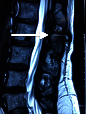
Methods: A 42-year-old female patient was attending a cesarean section eight years earlier to her consult. In the cesarean section, she received regional epidural anesthesia, and the main complaint was low back pain, specifically between the spinous processes L2 and L3. The somatic pain had been presenting intermittently for eight years. The sagittal section of magnetic resonance imaging of the lumbar spine showed a "golf club" image from the midline to the laminae of L2 and L3 with the subcutaneous tissue.
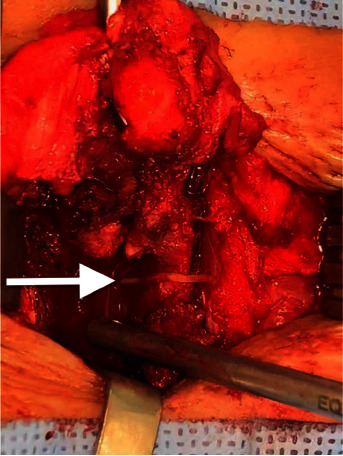
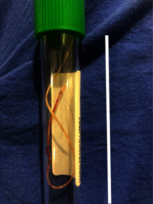
Results: A small right hemilaminectomy was performed to remove the complete catheter, which did not adhere, but was coiled in the S-shape. The catheter was trapped between the left facets of L2 and L3 laterally than through the midline. Several risk factors and therapeutic procedures have been proposed.
Conclusion: In a systematic review, 24 articles were reported on this specific issue. No surgical procedure and follow-up were informed by 8 authors. Surgical remotion by laminectomy was used in 9 articles, surgical explanation by skin incision was reported by 4 authors, and remotion by endoscopy was reported in 1 article. Two articles not reported solution.

 求助内容:
求助内容: 应助结果提醒方式:


