{"title":"细胞凋亡、自噬细胞死亡和坏死凋亡:牛黄体退化中不同类型的程序性细胞死亡。","authors":"Takuo Hojo, Dariusz J Skarzynski, Kiyoshi Okuda","doi":"10.1262/jrd.2022-097","DOIUrl":null,"url":null,"abstract":"<p><p>In mammals, the corpus luteum (CL) is a transient organ that secretes progesterone (P4). In the absence of pregnancy, the CL undergoes regression (luteolysis), which is a crucial preparation step for the next estrous cycle. Luteolysis, initiated by uterine prostaglandin F<sub>2α</sub> (PGF) in cattle, is usually divided into two phases, namely functional luteolysis characterized by a decline in P4 concentration and structural luteolysis characterized by the elimination of luteal tissues from the ovary. Programmed cell death (PCD) of luteal cells, including luteal steroidogenic cells (LSCs) and luteal endothelial cells (LECs), plays a crucial role in structural luteolysis. The main types of PCD are caspase-dependent apoptosis (type 1), autophagic cell death (ACD) via the autophagy-related gene (ATG) family (type 2), and receptor-interacting protein kinase (RIPK)-dependent programmed necrosis (necroptosis, type 3). However, these PCD signaling pathways are not completely independent and interact with each other. Over the past several decades, most studies on luteolysis have focused on apoptosis as the principal mode of bovine luteal cell death. Recently, ATG family members were reported to be expressed in bovine CL, and their levels increased during luteolysis. Furthermore, the expression of RIPKs, which are crucial mediators of necroptosis, is reported to increase in bovine CL during luteolysis and is upregulated by pro-inflammatory cytokines in bovine LSCs and LECs. Therefore, apoptosis, ACD, and necroptosis may contribute to bovine CL regression. In this article, we present the recent findings regarding the mechanisms of the three main types of PCD and the contribution of these mechanisms to luteolysis.</p>","PeriodicalId":16942,"journal":{"name":"Journal of Reproduction and Development","volume":"68 6","pages":"355-360"},"PeriodicalIF":1.9000,"publicationDate":"2022-12-19","publicationTypes":"Journal Article","fieldsOfStudy":null,"isOpenAccess":false,"openAccessPdf":"https://ftp.ncbi.nlm.nih.gov/pub/pmc/oa_pdf/40/31/jrd-68-355.PMC9792655.pdf","citationCount":"2","resultStr":"{\"title\":\"Apoptosis, autophagic cell death, and necroptosis: different types of programmed cell death in bovine corpus luteum regression.\",\"authors\":\"Takuo Hojo, Dariusz J Skarzynski, Kiyoshi Okuda\",\"doi\":\"10.1262/jrd.2022-097\",\"DOIUrl\":null,\"url\":null,\"abstract\":\"<p><p>In mammals, the corpus luteum (CL) is a transient organ that secretes progesterone (P4). In the absence of pregnancy, the CL undergoes regression (luteolysis), which is a crucial preparation step for the next estrous cycle. Luteolysis, initiated by uterine prostaglandin F<sub>2α</sub> (PGF) in cattle, is usually divided into two phases, namely functional luteolysis characterized by a decline in P4 concentration and structural luteolysis characterized by the elimination of luteal tissues from the ovary. Programmed cell death (PCD) of luteal cells, including luteal steroidogenic cells (LSCs) and luteal endothelial cells (LECs), plays a crucial role in structural luteolysis. The main types of PCD are caspase-dependent apoptosis (type 1), autophagic cell death (ACD) via the autophagy-related gene (ATG) family (type 2), and receptor-interacting protein kinase (RIPK)-dependent programmed necrosis (necroptosis, type 3). However, these PCD signaling pathways are not completely independent and interact with each other. Over the past several decades, most studies on luteolysis have focused on apoptosis as the principal mode of bovine luteal cell death. Recently, ATG family members were reported to be expressed in bovine CL, and their levels increased during luteolysis. Furthermore, the expression of RIPKs, which are crucial mediators of necroptosis, is reported to increase in bovine CL during luteolysis and is upregulated by pro-inflammatory cytokines in bovine LSCs and LECs. Therefore, apoptosis, ACD, and necroptosis may contribute to bovine CL regression. In this article, we present the recent findings regarding the mechanisms of the three main types of PCD and the contribution of these mechanisms to luteolysis.</p>\",\"PeriodicalId\":16942,\"journal\":{\"name\":\"Journal of Reproduction and Development\",\"volume\":\"68 6\",\"pages\":\"355-360\"},\"PeriodicalIF\":1.9000,\"publicationDate\":\"2022-12-19\",\"publicationTypes\":\"Journal Article\",\"fieldsOfStudy\":null,\"isOpenAccess\":false,\"openAccessPdf\":\"https://ftp.ncbi.nlm.nih.gov/pub/pmc/oa_pdf/40/31/jrd-68-355.PMC9792655.pdf\",\"citationCount\":\"2\",\"resultStr\":null,\"platform\":\"Semanticscholar\",\"paperid\":null,\"PeriodicalName\":\"Journal of Reproduction and Development\",\"FirstCategoryId\":\"99\",\"ListUrlMain\":\"https://doi.org/10.1262/jrd.2022-097\",\"RegionNum\":4,\"RegionCategory\":\"生物学\",\"ArticlePicture\":[],\"TitleCN\":null,\"AbstractTextCN\":null,\"PMCID\":null,\"EPubDate\":\"\",\"PubModel\":\"\",\"JCR\":\"Q2\",\"JCRName\":\"AGRICULTURE, DAIRY & ANIMAL SCIENCE\",\"Score\":null,\"Total\":0}","platform":"Semanticscholar","paperid":null,"PeriodicalName":"Journal of Reproduction and Development","FirstCategoryId":"99","ListUrlMain":"https://doi.org/10.1262/jrd.2022-097","RegionNum":4,"RegionCategory":"生物学","ArticlePicture":[],"TitleCN":null,"AbstractTextCN":null,"PMCID":null,"EPubDate":"","PubModel":"","JCR":"Q2","JCRName":"AGRICULTURE, DAIRY & ANIMAL SCIENCE","Score":null,"Total":0}
Apoptosis, autophagic cell death, and necroptosis: different types of programmed cell death in bovine corpus luteum regression.
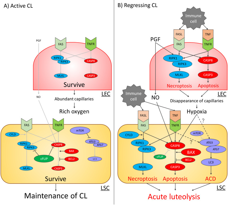
In mammals, the corpus luteum (CL) is a transient organ that secretes progesterone (P4). In the absence of pregnancy, the CL undergoes regression (luteolysis), which is a crucial preparation step for the next estrous cycle. Luteolysis, initiated by uterine prostaglandin F2α (PGF) in cattle, is usually divided into two phases, namely functional luteolysis characterized by a decline in P4 concentration and structural luteolysis characterized by the elimination of luteal tissues from the ovary. Programmed cell death (PCD) of luteal cells, including luteal steroidogenic cells (LSCs) and luteal endothelial cells (LECs), plays a crucial role in structural luteolysis. The main types of PCD are caspase-dependent apoptosis (type 1), autophagic cell death (ACD) via the autophagy-related gene (ATG) family (type 2), and receptor-interacting protein kinase (RIPK)-dependent programmed necrosis (necroptosis, type 3). However, these PCD signaling pathways are not completely independent and interact with each other. Over the past several decades, most studies on luteolysis have focused on apoptosis as the principal mode of bovine luteal cell death. Recently, ATG family members were reported to be expressed in bovine CL, and their levels increased during luteolysis. Furthermore, the expression of RIPKs, which are crucial mediators of necroptosis, is reported to increase in bovine CL during luteolysis and is upregulated by pro-inflammatory cytokines in bovine LSCs and LECs. Therefore, apoptosis, ACD, and necroptosis may contribute to bovine CL regression. In this article, we present the recent findings regarding the mechanisms of the three main types of PCD and the contribution of these mechanisms to luteolysis.
期刊介绍:
Journal of Reproduction and Development (JRD) is the
official journal of the Society for Reproduction and Development,
published bimonthly, and welcomes original articles. JRD
provides free full-text access of all the published articles on
the web. The functions of the journal are managed by Editorial
Board Members, such as the Editor-in-Chief, Co-Editor-inChief, Managing Editors and Editors. All manuscripts are
peer-reviewed critically by two or more reviewers. Acceptance
is based on scientific content and presentation of the materials.
The Editors select reviewers and correspond with authors. Final
decisions about acceptance or rejection of manuscripts are made
by the Editor-in-Chief and Co-Editor-in-Chief.
 求助内容:
求助内容: 应助结果提醒方式:


