Jasmin Wächter , Matthew J. Shannon , Alexander G. Beristain
{"title":"人滋养细胞中metzincin景观的转录组作图","authors":"Jasmin Wächter , Matthew J. Shannon , Alexander G. Beristain","doi":"10.1016/j.gep.2022.119283","DOIUrl":null,"url":null,"abstract":"<div><p><span><span>The metzincin family of metalloproteases<span><span><span> coordinates tissue developmental processes through regulation of growth factor availability, receptor signaling, and cell-cell/cell-matrix adhesion. While roles for select metzincins in controlling trophoblast functions in human placental development have been described, a comprehensive understanding of metzincin dynamics during trophoblast differentiation is lacking. To address this knowledge gap, single cell </span>transcriptomic<span> datasets derived from first trimester<span> chorionic villi and decidua were used to decipher metzincin expression profiles and kinetics in diverse cell types within the utero-placental interface. Further, specific protease-substrate interactions within progenitor trophoblasts were examined to better define the progenitor niche. Within the uterine-placental compartment, 43 metzincin proteases were expressed across 15 cell-type clusters. Metzincin subgroups expressed in placental trophoblasts, placental mesenchymal cells, uterine stromal, and </span></span></span>immune cells<span> included multiple matrix metalloproteases (MMPs), a disintegrin and metalloproteases (ADAMs), a disintegrin and metalloproteases with </span></span></span>thrombospondin<span> repeats (ADAMTSs), pappalysins, and astacins<span><span>. Within the trophoblast compartment, eight distinct trophoblasts states were identified: four cytotrophoblast (CTB), one </span>syncytiotrophoblast precursor (SCTp), two column CTB (cCTB), and one extravillous trophoblast (EVT). Within these states 7 MMP, 8 ADAM, 4 ADAMTS, 2 pappalysin, and 3 astacin proteases were expressed. Cell trajectory modeling shows that expression of most (19/24) metzincins increase during EVT differentiation, though expression of select metalloproteases increase along the villous pathway. Eleven metzincins (</span></span></span><span><em>ADAM10</em></span>, <em>-17</em>, <span><em>MMP14</em></span>, <em>-15, -19, -23B</em>, <span><em>ADAMTS1</em><em>, -6, -19, TLL-1, -2</em></span>) showed enrichment within CTB progenitors, and analysis of metzincin-substrate interactions identified ∼150 substrates and binding partners, including <em>FBN2</em> as an <em>ADAMTS6</em>-specific substrate. Together, this work characterizes the metzincin landscape in human first trimester trophoblasts and establishes insight into the roles specific proteases perform within distinct trophoblast niches and across trophoblast differentiation. This resource serves as a guide for future investigations into the roles of metzincin proteases in human placental development.</p></div>","PeriodicalId":55598,"journal":{"name":"Gene Expression Patterns","volume":"46 ","pages":"Article 119283"},"PeriodicalIF":1.0000,"publicationDate":"2022-12-01","publicationTypes":"Journal Article","fieldsOfStudy":null,"isOpenAccess":false,"openAccessPdf":"","citationCount":"0","resultStr":"{\"title\":\"Transcriptomic mapping of the metzincin landscape in human trophoblasts\",\"authors\":\"Jasmin Wächter , Matthew J. Shannon , Alexander G. Beristain\",\"doi\":\"10.1016/j.gep.2022.119283\",\"DOIUrl\":null,\"url\":null,\"abstract\":\"<div><p><span><span>The metzincin family of metalloproteases<span><span><span> coordinates tissue developmental processes through regulation of growth factor availability, receptor signaling, and cell-cell/cell-matrix adhesion. While roles for select metzincins in controlling trophoblast functions in human placental development have been described, a comprehensive understanding of metzincin dynamics during trophoblast differentiation is lacking. To address this knowledge gap, single cell </span>transcriptomic<span> datasets derived from first trimester<span> chorionic villi and decidua were used to decipher metzincin expression profiles and kinetics in diverse cell types within the utero-placental interface. Further, specific protease-substrate interactions within progenitor trophoblasts were examined to better define the progenitor niche. Within the uterine-placental compartment, 43 metzincin proteases were expressed across 15 cell-type clusters. Metzincin subgroups expressed in placental trophoblasts, placental mesenchymal cells, uterine stromal, and </span></span></span>immune cells<span> included multiple matrix metalloproteases (MMPs), a disintegrin and metalloproteases (ADAMs), a disintegrin and metalloproteases with </span></span></span>thrombospondin<span> repeats (ADAMTSs), pappalysins, and astacins<span><span>. Within the trophoblast compartment, eight distinct trophoblasts states were identified: four cytotrophoblast (CTB), one </span>syncytiotrophoblast precursor (SCTp), two column CTB (cCTB), and one extravillous trophoblast (EVT). Within these states 7 MMP, 8 ADAM, 4 ADAMTS, 2 pappalysin, and 3 astacin proteases were expressed. Cell trajectory modeling shows that expression of most (19/24) metzincins increase during EVT differentiation, though expression of select metalloproteases increase along the villous pathway. Eleven metzincins (</span></span></span><span><em>ADAM10</em></span>, <em>-17</em>, <span><em>MMP14</em></span>, <em>-15, -19, -23B</em>, <span><em>ADAMTS1</em><em>, -6, -19, TLL-1, -2</em></span>) showed enrichment within CTB progenitors, and analysis of metzincin-substrate interactions identified ∼150 substrates and binding partners, including <em>FBN2</em> as an <em>ADAMTS6</em>-specific substrate. Together, this work characterizes the metzincin landscape in human first trimester trophoblasts and establishes insight into the roles specific proteases perform within distinct trophoblast niches and across trophoblast differentiation. This resource serves as a guide for future investigations into the roles of metzincin proteases in human placental development.</p></div>\",\"PeriodicalId\":55598,\"journal\":{\"name\":\"Gene Expression Patterns\",\"volume\":\"46 \",\"pages\":\"Article 119283\"},\"PeriodicalIF\":1.0000,\"publicationDate\":\"2022-12-01\",\"publicationTypes\":\"Journal Article\",\"fieldsOfStudy\":null,\"isOpenAccess\":false,\"openAccessPdf\":\"\",\"citationCount\":\"0\",\"resultStr\":null,\"platform\":\"Semanticscholar\",\"paperid\":null,\"PeriodicalName\":\"Gene Expression Patterns\",\"FirstCategoryId\":\"99\",\"ListUrlMain\":\"https://www.sciencedirect.com/science/article/pii/S1567133X22000539\",\"RegionNum\":4,\"RegionCategory\":\"生物学\",\"ArticlePicture\":[],\"TitleCN\":null,\"AbstractTextCN\":null,\"PMCID\":null,\"EPubDate\":\"\",\"PubModel\":\"\",\"JCR\":\"Q4\",\"JCRName\":\"DEVELOPMENTAL BIOLOGY\",\"Score\":null,\"Total\":0}","platform":"Semanticscholar","paperid":null,"PeriodicalName":"Gene Expression Patterns","FirstCategoryId":"99","ListUrlMain":"https://www.sciencedirect.com/science/article/pii/S1567133X22000539","RegionNum":4,"RegionCategory":"生物学","ArticlePicture":[],"TitleCN":null,"AbstractTextCN":null,"PMCID":null,"EPubDate":"","PubModel":"","JCR":"Q4","JCRName":"DEVELOPMENTAL BIOLOGY","Score":null,"Total":0}
Transcriptomic mapping of the metzincin landscape in human trophoblasts
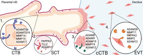
The metzincin family of metalloproteases coordinates tissue developmental processes through regulation of growth factor availability, receptor signaling, and cell-cell/cell-matrix adhesion. While roles for select metzincins in controlling trophoblast functions in human placental development have been described, a comprehensive understanding of metzincin dynamics during trophoblast differentiation is lacking. To address this knowledge gap, single cell transcriptomic datasets derived from first trimester chorionic villi and decidua were used to decipher metzincin expression profiles and kinetics in diverse cell types within the utero-placental interface. Further, specific protease-substrate interactions within progenitor trophoblasts were examined to better define the progenitor niche. Within the uterine-placental compartment, 43 metzincin proteases were expressed across 15 cell-type clusters. Metzincin subgroups expressed in placental trophoblasts, placental mesenchymal cells, uterine stromal, and immune cells included multiple matrix metalloproteases (MMPs), a disintegrin and metalloproteases (ADAMs), a disintegrin and metalloproteases with thrombospondin repeats (ADAMTSs), pappalysins, and astacins. Within the trophoblast compartment, eight distinct trophoblasts states were identified: four cytotrophoblast (CTB), one syncytiotrophoblast precursor (SCTp), two column CTB (cCTB), and one extravillous trophoblast (EVT). Within these states 7 MMP, 8 ADAM, 4 ADAMTS, 2 pappalysin, and 3 astacin proteases were expressed. Cell trajectory modeling shows that expression of most (19/24) metzincins increase during EVT differentiation, though expression of select metalloproteases increase along the villous pathway. Eleven metzincins (ADAM10, -17, MMP14, -15, -19, -23B, ADAMTS1, -6, -19, TLL-1, -2) showed enrichment within CTB progenitors, and analysis of metzincin-substrate interactions identified ∼150 substrates and binding partners, including FBN2 as an ADAMTS6-specific substrate. Together, this work characterizes the metzincin landscape in human first trimester trophoblasts and establishes insight into the roles specific proteases perform within distinct trophoblast niches and across trophoblast differentiation. This resource serves as a guide for future investigations into the roles of metzincin proteases in human placental development.
期刊介绍:
Gene Expression Patterns is devoted to the rapid publication of high quality studies of gene expression in development. Studies using cell culture are also suitable if clearly relevant to development, e.g., analysis of key regulatory genes or of gene sets in the maintenance or differentiation of stem cells. Key areas of interest include:
-In-situ studies such as expression patterns of important or interesting genes at all levels, including transcription and protein expression
-Temporal studies of large gene sets during development
-Transgenic studies to study cell lineage in tissue formation
 求助内容:
求助内容: 应助结果提醒方式:


