{"title":"角膜缘缝合大鼠眼压模型巩膜的区域差异。","authors":"Mingfang Xia, Endong Zhang, Fei Yao, Zhaohua Xia, Mingmin Zhou, Xufang Ran, Xiaobo Xia","doi":"10.1186/s40662-022-00319-w","DOIUrl":null,"url":null,"abstract":"<p><strong>Purpose: </strong>To describe the regional differences of the sclera in ocular hypertension (OHT) models with the inappropriate extension of the ocular axis.</p><p><strong>Methods: </strong>To discover the regional differences of the sclera at the early stage, OHT models were established using circumlimbal suture (CS) or sclerosant injection (SI). Axial length (AL) was measured by ultrasound and magnetic resonance imaging. The glaucoma-associated distinction was determined by intraocular pressure (IOP) and retrograde tracing of retinal ganglion cells (RGCs). The central thickness of the ganglion cell complex (GCC) was measured by optical coherence tomography. RGCs and collagen fibrils were detected using a transmission electron microscope, furthermore, anti-alpha smooth muscle actin (αSMA) was determined in the early stage after the operation.</p><p><strong>Results: </strong>Compared with the control group, the eyes in OHT models showed an increased IOP (P < 0.001 in the CS group, P = 0.001 in the SI group), growing AL (P = 0.026 in the CS group, P = 0.043 in the SI group), reduction of central RGCs (P < 0.001 in the CS group, P = 0.017 in the SI group), thinning central GCC (P < 0.001 in the CS group), and a distinctive expression of αSMA in the central sclera in the early 4-week stage after the operation (P = 0.002 in the CS group). Compared with the SI group, the eye in the CS group showed a significantly increased AL (7.1 ± 0.4 mm, P = 0.031), reduction of central RGCs (2121.1 ± 87.2 cells/mm<sup>2</sup>, P = 0.001), thinning central GCC (71.4 ± 0.8 pixels, P = 0.015), and a distinctive expression of αSMA (P = 0.005). Additionally, ultrastructural changes in RGCs, scleral collagen fibers, and collagen crimp were observed in the different regions. Increased collagen volume fraction in the posterior segment of the eyeball wall (30.2 ± 3.1%, P = 0.022) was observed by MASSON staining in the CS group.</p><p><strong>Conclusion: </strong>Regional differences of the sclera in the ocular hypertensive rat model induced by CS may provide a reference for further treatment of scleral-related eye disorders.</p>","PeriodicalId":12194,"journal":{"name":"Eye and Vision","volume":null,"pages":null},"PeriodicalIF":4.1000,"publicationDate":"2023-01-04","publicationTypes":"Journal Article","fieldsOfStudy":null,"isOpenAccess":false,"openAccessPdf":"https://www.ncbi.nlm.nih.gov/pmc/articles/PMC9811703/pdf/","citationCount":"0","resultStr":"{\"title\":\"Regional differences of the sclera in the ocular hypertensive rat model induced by circumlimbal suture.\",\"authors\":\"Mingfang Xia, Endong Zhang, Fei Yao, Zhaohua Xia, Mingmin Zhou, Xufang Ran, Xiaobo Xia\",\"doi\":\"10.1186/s40662-022-00319-w\",\"DOIUrl\":null,\"url\":null,\"abstract\":\"<p><strong>Purpose: </strong>To describe the regional differences of the sclera in ocular hypertension (OHT) models with the inappropriate extension of the ocular axis.</p><p><strong>Methods: </strong>To discover the regional differences of the sclera at the early stage, OHT models were established using circumlimbal suture (CS) or sclerosant injection (SI). Axial length (AL) was measured by ultrasound and magnetic resonance imaging. The glaucoma-associated distinction was determined by intraocular pressure (IOP) and retrograde tracing of retinal ganglion cells (RGCs). The central thickness of the ganglion cell complex (GCC) was measured by optical coherence tomography. RGCs and collagen fibrils were detected using a transmission electron microscope, furthermore, anti-alpha smooth muscle actin (αSMA) was determined in the early stage after the operation.</p><p><strong>Results: </strong>Compared with the control group, the eyes in OHT models showed an increased IOP (P < 0.001 in the CS group, P = 0.001 in the SI group), growing AL (P = 0.026 in the CS group, P = 0.043 in the SI group), reduction of central RGCs (P < 0.001 in the CS group, P = 0.017 in the SI group), thinning central GCC (P < 0.001 in the CS group), and a distinctive expression of αSMA in the central sclera in the early 4-week stage after the operation (P = 0.002 in the CS group). Compared with the SI group, the eye in the CS group showed a significantly increased AL (7.1 ± 0.4 mm, P = 0.031), reduction of central RGCs (2121.1 ± 87.2 cells/mm<sup>2</sup>, P = 0.001), thinning central GCC (71.4 ± 0.8 pixels, P = 0.015), and a distinctive expression of αSMA (P = 0.005). Additionally, ultrastructural changes in RGCs, scleral collagen fibers, and collagen crimp were observed in the different regions. Increased collagen volume fraction in the posterior segment of the eyeball wall (30.2 ± 3.1%, P = 0.022) was observed by MASSON staining in the CS group.</p><p><strong>Conclusion: </strong>Regional differences of the sclera in the ocular hypertensive rat model induced by CS may provide a reference for further treatment of scleral-related eye disorders.</p>\",\"PeriodicalId\":12194,\"journal\":{\"name\":\"Eye and Vision\",\"volume\":null,\"pages\":null},\"PeriodicalIF\":4.1000,\"publicationDate\":\"2023-01-04\",\"publicationTypes\":\"Journal Article\",\"fieldsOfStudy\":null,\"isOpenAccess\":false,\"openAccessPdf\":\"https://www.ncbi.nlm.nih.gov/pmc/articles/PMC9811703/pdf/\",\"citationCount\":\"0\",\"resultStr\":null,\"platform\":\"Semanticscholar\",\"paperid\":null,\"PeriodicalName\":\"Eye and Vision\",\"FirstCategoryId\":\"3\",\"ListUrlMain\":\"https://doi.org/10.1186/s40662-022-00319-w\",\"RegionNum\":1,\"RegionCategory\":\"医学\",\"ArticlePicture\":[],\"TitleCN\":null,\"AbstractTextCN\":null,\"PMCID\":null,\"EPubDate\":\"\",\"PubModel\":\"\",\"JCR\":\"Q1\",\"JCRName\":\"OPHTHALMOLOGY\",\"Score\":null,\"Total\":0}","platform":"Semanticscholar","paperid":null,"PeriodicalName":"Eye and Vision","FirstCategoryId":"3","ListUrlMain":"https://doi.org/10.1186/s40662-022-00319-w","RegionNum":1,"RegionCategory":"医学","ArticlePicture":[],"TitleCN":null,"AbstractTextCN":null,"PMCID":null,"EPubDate":"","PubModel":"","JCR":"Q1","JCRName":"OPHTHALMOLOGY","Score":null,"Total":0}
Regional differences of the sclera in the ocular hypertensive rat model induced by circumlimbal suture.
Purpose: To describe the regional differences of the sclera in ocular hypertension (OHT) models with the inappropriate extension of the ocular axis.
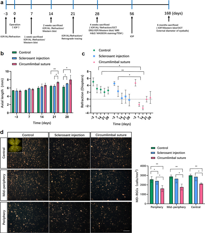
Methods: To discover the regional differences of the sclera at the early stage, OHT models were established using circumlimbal suture (CS) or sclerosant injection (SI). Axial length (AL) was measured by ultrasound and magnetic resonance imaging. The glaucoma-associated distinction was determined by intraocular pressure (IOP) and retrograde tracing of retinal ganglion cells (RGCs). The central thickness of the ganglion cell complex (GCC) was measured by optical coherence tomography. RGCs and collagen fibrils were detected using a transmission electron microscope, furthermore, anti-alpha smooth muscle actin (αSMA) was determined in the early stage after the operation.
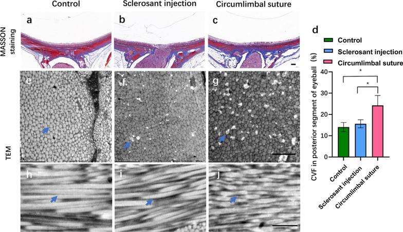
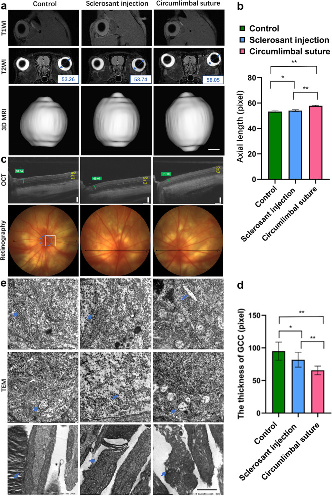
Results: Compared with the control group, the eyes in OHT models showed an increased IOP (P < 0.001 in the CS group, P = 0.001 in the SI group), growing AL (P = 0.026 in the CS group, P = 0.043 in the SI group), reduction of central RGCs (P < 0.001 in the CS group, P = 0.017 in the SI group), thinning central GCC (P < 0.001 in the CS group), and a distinctive expression of αSMA in the central sclera in the early 4-week stage after the operation (P = 0.002 in the CS group). Compared with the SI group, the eye in the CS group showed a significantly increased AL (7.1 ± 0.4 mm, P = 0.031), reduction of central RGCs (2121.1 ± 87.2 cells/mm2, P = 0.001), thinning central GCC (71.4 ± 0.8 pixels, P = 0.015), and a distinctive expression of αSMA (P = 0.005). Additionally, ultrastructural changes in RGCs, scleral collagen fibers, and collagen crimp were observed in the different regions. Increased collagen volume fraction in the posterior segment of the eyeball wall (30.2 ± 3.1%, P = 0.022) was observed by MASSON staining in the CS group.
Conclusion: Regional differences of the sclera in the ocular hypertensive rat model induced by CS may provide a reference for further treatment of scleral-related eye disorders.
期刊介绍:
Eye and Vision is an open access, peer-reviewed journal for ophthalmologists and visual science specialists. It welcomes research articles, reviews, methodologies, commentaries, case reports, perspectives and short reports encompassing all aspects of eye and vision. Topics of interest include but are not limited to: current developments of theoretical, experimental and clinical investigations in ophthalmology, optometry and vision science which focus on novel and high-impact findings on central issues pertaining to biology, pathophysiology and etiology of eye diseases as well as advances in diagnostic techniques, surgical treatment, instrument updates, the latest drug findings, results of clinical trials and research findings. It aims to provide ophthalmologists and visual science specialists with the latest developments in theoretical, experimental and clinical investigations in eye and vision.
 求助内容:
求助内容: 应助结果提醒方式:


