你的诊断是什么?
IF 16.4
1区 化学
Q1 CHEMISTRY, MULTIDISCIPLINARY
引用次数: 0
摘要
本文章由计算机程序翻译,如有差异,请以英文原文为准。
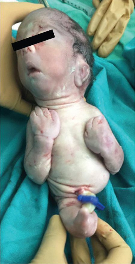
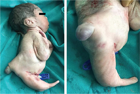
What is your diagnosis?
A 25-year-old primigravida, was admitted to the antenatal ward at 32 weeks gestation with decreased fetal movements. The patient lived in a remote hilly region and did not go for antenatal checkups because of the non-availability of transport due to the lockdown imposed during the coronavirus pandemic. The patient did not undergo an anomaly scan in the first or second trimester. The present pregnancy was conceived spontaneously, without any history of ovulation induction. There was no history of consanguineous marriage. There was no history of teratogenic drug exposure in the antenatal period. The patient did not have any risk factors for gestational diabetes, body mass index was 22.6 kg/m 2 , and family history was not significant. Blood sugar profile was normal after admission, and during the intrapartum and postpartum periods while hemoglobinA1c was normal at 5.8%. On examination, the fundal height corresponded to 26 weeks, and fetal parts were palpable superficially, suggesting decreased liquor and fetal growth restriction (FGR). The ultrasound showed a single live fetus in breech presentation, corresponding to gestational age 32 weeks with severe FGR, abdominal circumference less than the third centile, biparietal diameter and head circumference at the fifth centile and femur length at the tenth centile with placenta praevia and almost absent liquor. Due to grossly decreased liquor, the radiologist could not comment on fetal anatomy at this gestation. A Doppler study of the umbilical arteries suggested reversed end-diastolic flow with brain sparing effect. Cardiotocography was suggestive of prolonged late decelerations. After discussion with the parents, the patient was taken for lower segment caesarean section because of primigravida with placenta praevia, breech presentation and Stage 4 FGR with high suspicion of fetal acidosis (1).
求助全文
通过发布文献求助,成功后即可免费获取论文全文。
去求助
来源期刊

Accounts of Chemical Research
化学-化学综合
CiteScore
31.40
自引率
1.10%
发文量
312
审稿时长
2 months
期刊介绍:
Accounts of Chemical Research presents short, concise and critical articles offering easy-to-read overviews of basic research and applications in all areas of chemistry and biochemistry. These short reviews focus on research from the author’s own laboratory and are designed to teach the reader about a research project. In addition, Accounts of Chemical Research publishes commentaries that give an informed opinion on a current research problem. Special Issues online are devoted to a single topic of unusual activity and significance.
Accounts of Chemical Research replaces the traditional article abstract with an article "Conspectus." These entries synopsize the research affording the reader a closer look at the content and significance of an article. Through this provision of a more detailed description of the article contents, the Conspectus enhances the article's discoverability by search engines and the exposure for the research.
 求助内容:
求助内容: 应助结果提醒方式:


