Liwen Zhang, Runzhi Guo, Baohua Xu, Yue Wang, Weiran Li
{"title":"三种不同微支抗系统拔牙患者上颌牙移动的三维评价:一项随机对照试验。","authors":"Liwen Zhang, Runzhi Guo, Baohua Xu, Yue Wang, Weiran Li","doi":"10.1186/s40510-022-00441-4","DOIUrl":null,"url":null,"abstract":"<p><strong>Objective: </strong>To compare the three-dimensional (3-D) movement of maxillary teeth in response to three common miniscrew anchorage systems in extraction patients with maxillary dentoalveolar protrusion.</p><p><strong>Materials and methods: </strong>The study employed a randomized controlled single-blinded design with three arms. Thirty extraction patients who required maximum anchorage to retract maxillary anterior teeth were included and randomly allocated into three treatment groups: space closure with direct miniscrew anchorage and low crimpable hooks (DL group), indirect miniscrew anchorage and low crimpable hooks (IL group), and direct miniscrew anchorage and high crimpable hooks (DH group). Cone beam computed tomography (CBCT) images of all included patients were obtained immediately before (T0) and after (T1) space closure. The outcomes were 3-D positional changes of maxillary central incisor, lateral incisor, canine, second premolar, and first molar. The repeated measures analysis of variance with post hoc LSD test was used to evaluate differences among groups.</p><p><strong>Results: </strong>A significant intrusion (- 1.34 mm; 95% CI, - 1.60 mm, 1.08 mm) and buccal (- 6.92°; 95% CI, - 8.67°, - 5.13°) and distal (4.90°; 95% CI, 3.75°, 6.04°) inclination of the maxillary first molars were observed in the DL group, compared to the other two groups. The mesial movement (- 0.40 mm; 95% CI, - 0.83 mm, - 0.03 mm) of the maxillary first molars was found in the IL group, while the DL (0.44 mm; 95% CI, 0.15 mm, 0.73 mm) and IL (0.62 mm; 95% CI, 0.28 mm, 0.96 mm) groups exhibited distal movement. In the DH group, the lingual inclination changes of maxillary central incisor (5.04°; 95% CI, 2.82°, 7.26°) were significantly lower, which is indicative of good lingual root torque control of the maxillary anterior teeth.</p><p><strong>Conclusion: </strong>Three miniscrew anchorage systems produced significantly different 3-D maxillary tooth movement. The maxillary first molars were significantly buccally and distally inclined and intruded in patients using direct miniscrew anchorages with low crimpable hooks. Direct miniscrew anchorages with high crimpable hooks could help to achieve better lingual root torque control of the maxillary incisors. Trial registration The trial was registered at www.chictr.org.cn (ChiCTR1900026960). Registered 27 October 2019.</p>","PeriodicalId":56071,"journal":{"name":"Progress in Orthodontics","volume":"23 1","pages":"46"},"PeriodicalIF":4.8000,"publicationDate":"2022-12-19","publicationTypes":"Journal Article","fieldsOfStudy":null,"isOpenAccess":false,"openAccessPdf":"https://www.ncbi.nlm.nih.gov/pmc/articles/PMC9760583/pdf/","citationCount":"1","resultStr":"{\"title\":\"Three-dimensional evaluation of maxillary tooth movement in extraction patients with three different miniscrew anchorage systems: a randomized controlled trial.\",\"authors\":\"Liwen Zhang, Runzhi Guo, Baohua Xu, Yue Wang, Weiran Li\",\"doi\":\"10.1186/s40510-022-00441-4\",\"DOIUrl\":null,\"url\":null,\"abstract\":\"<p><strong>Objective: </strong>To compare the three-dimensional (3-D) movement of maxillary teeth in response to three common miniscrew anchorage systems in extraction patients with maxillary dentoalveolar protrusion.</p><p><strong>Materials and methods: </strong>The study employed a randomized controlled single-blinded design with three arms. Thirty extraction patients who required maximum anchorage to retract maxillary anterior teeth were included and randomly allocated into three treatment groups: space closure with direct miniscrew anchorage and low crimpable hooks (DL group), indirect miniscrew anchorage and low crimpable hooks (IL group), and direct miniscrew anchorage and high crimpable hooks (DH group). Cone beam computed tomography (CBCT) images of all included patients were obtained immediately before (T0) and after (T1) space closure. The outcomes were 3-D positional changes of maxillary central incisor, lateral incisor, canine, second premolar, and first molar. The repeated measures analysis of variance with post hoc LSD test was used to evaluate differences among groups.</p><p><strong>Results: </strong>A significant intrusion (- 1.34 mm; 95% CI, - 1.60 mm, 1.08 mm) and buccal (- 6.92°; 95% CI, - 8.67°, - 5.13°) and distal (4.90°; 95% CI, 3.75°, 6.04°) inclination of the maxillary first molars were observed in the DL group, compared to the other two groups. The mesial movement (- 0.40 mm; 95% CI, - 0.83 mm, - 0.03 mm) of the maxillary first molars was found in the IL group, while the DL (0.44 mm; 95% CI, 0.15 mm, 0.73 mm) and IL (0.62 mm; 95% CI, 0.28 mm, 0.96 mm) groups exhibited distal movement. In the DH group, the lingual inclination changes of maxillary central incisor (5.04°; 95% CI, 2.82°, 7.26°) were significantly lower, which is indicative of good lingual root torque control of the maxillary anterior teeth.</p><p><strong>Conclusion: </strong>Three miniscrew anchorage systems produced significantly different 3-D maxillary tooth movement. The maxillary first molars were significantly buccally and distally inclined and intruded in patients using direct miniscrew anchorages with low crimpable hooks. Direct miniscrew anchorages with high crimpable hooks could help to achieve better lingual root torque control of the maxillary incisors. Trial registration The trial was registered at www.chictr.org.cn (ChiCTR1900026960). Registered 27 October 2019.</p>\",\"PeriodicalId\":56071,\"journal\":{\"name\":\"Progress in Orthodontics\",\"volume\":\"23 1\",\"pages\":\"46\"},\"PeriodicalIF\":4.8000,\"publicationDate\":\"2022-12-19\",\"publicationTypes\":\"Journal Article\",\"fieldsOfStudy\":null,\"isOpenAccess\":false,\"openAccessPdf\":\"https://www.ncbi.nlm.nih.gov/pmc/articles/PMC9760583/pdf/\",\"citationCount\":\"1\",\"resultStr\":null,\"platform\":\"Semanticscholar\",\"paperid\":null,\"PeriodicalName\":\"Progress in Orthodontics\",\"FirstCategoryId\":\"3\",\"ListUrlMain\":\"https://doi.org/10.1186/s40510-022-00441-4\",\"RegionNum\":2,\"RegionCategory\":\"医学\",\"ArticlePicture\":[],\"TitleCN\":null,\"AbstractTextCN\":null,\"PMCID\":null,\"EPubDate\":\"\",\"PubModel\":\"\",\"JCR\":\"Q1\",\"JCRName\":\"Dentistry\",\"Score\":null,\"Total\":0}","platform":"Semanticscholar","paperid":null,"PeriodicalName":"Progress in Orthodontics","FirstCategoryId":"3","ListUrlMain":"https://doi.org/10.1186/s40510-022-00441-4","RegionNum":2,"RegionCategory":"医学","ArticlePicture":[],"TitleCN":null,"AbstractTextCN":null,"PMCID":null,"EPubDate":"","PubModel":"","JCR":"Q1","JCRName":"Dentistry","Score":null,"Total":0}
Three-dimensional evaluation of maxillary tooth movement in extraction patients with three different miniscrew anchorage systems: a randomized controlled trial.
Objective: To compare the three-dimensional (3-D) movement of maxillary teeth in response to three common miniscrew anchorage systems in extraction patients with maxillary dentoalveolar protrusion.
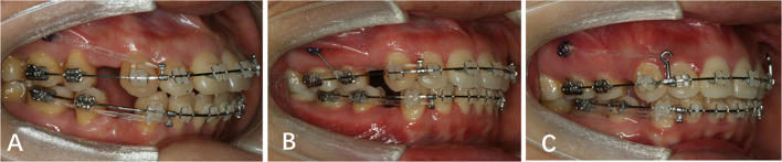
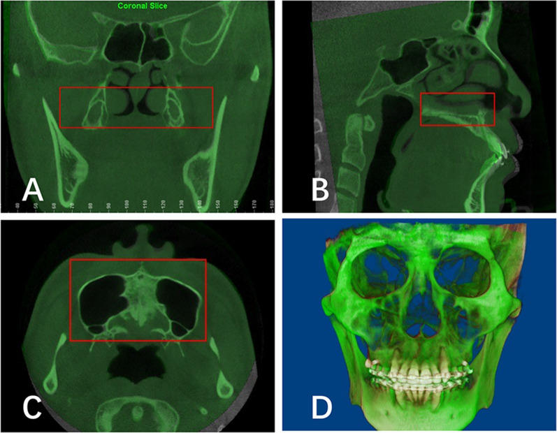
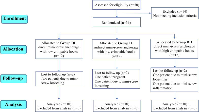
Materials and methods: The study employed a randomized controlled single-blinded design with three arms. Thirty extraction patients who required maximum anchorage to retract maxillary anterior teeth were included and randomly allocated into three treatment groups: space closure with direct miniscrew anchorage and low crimpable hooks (DL group), indirect miniscrew anchorage and low crimpable hooks (IL group), and direct miniscrew anchorage and high crimpable hooks (DH group). Cone beam computed tomography (CBCT) images of all included patients were obtained immediately before (T0) and after (T1) space closure. The outcomes were 3-D positional changes of maxillary central incisor, lateral incisor, canine, second premolar, and first molar. The repeated measures analysis of variance with post hoc LSD test was used to evaluate differences among groups.
Results: A significant intrusion (- 1.34 mm; 95% CI, - 1.60 mm, 1.08 mm) and buccal (- 6.92°; 95% CI, - 8.67°, - 5.13°) and distal (4.90°; 95% CI, 3.75°, 6.04°) inclination of the maxillary first molars were observed in the DL group, compared to the other two groups. The mesial movement (- 0.40 mm; 95% CI, - 0.83 mm, - 0.03 mm) of the maxillary first molars was found in the IL group, while the DL (0.44 mm; 95% CI, 0.15 mm, 0.73 mm) and IL (0.62 mm; 95% CI, 0.28 mm, 0.96 mm) groups exhibited distal movement. In the DH group, the lingual inclination changes of maxillary central incisor (5.04°; 95% CI, 2.82°, 7.26°) were significantly lower, which is indicative of good lingual root torque control of the maxillary anterior teeth.
Conclusion: Three miniscrew anchorage systems produced significantly different 3-D maxillary tooth movement. The maxillary first molars were significantly buccally and distally inclined and intruded in patients using direct miniscrew anchorages with low crimpable hooks. Direct miniscrew anchorages with high crimpable hooks could help to achieve better lingual root torque control of the maxillary incisors. Trial registration The trial was registered at www.chictr.org.cn (ChiCTR1900026960). Registered 27 October 2019.
期刊介绍:
Progress in Orthodontics is a fully open access, international journal owned by the Italian Society of Orthodontics and published under the brand SpringerOpen. The Society is currently covering all publication costs so there are no article processing charges for authors.
It is a premier journal of international scope that fosters orthodontic research, including both basic research and development of innovative clinical techniques, with an emphasis on the following areas:
• Mechanisms to improve orthodontics
• Clinical studies and control animal studies
• Orthodontics and genetics, genomics
• Temporomandibular joint (TMJ) control clinical trials
• Efficacy of orthodontic appliances and animal models
• Systematic reviews and meta analyses
• Mechanisms to speed orthodontic treatment
Progress in Orthodontics will consider for publication only meritorious and original contributions. These may be:
• Original articles reporting the findings of clinical trials, clinically relevant basic scientific investigations, or novel therapeutic or diagnostic systems
• Review articles on current topics
• Articles on novel techniques and clinical tools
• Articles of contemporary interest
 求助内容:
求助内容: 应助结果提醒方式:


