{"title":"应用基于体素的单例形态计量学分析儿童交替性偏瘫的脑结构变化。","authors":"Elly Arizono, Noriko Sato, Yoko Shigemoto, Yukio Kimura, Emiko Chiba, Hiroyuki Maki, Hiroshi Matsuda, Eri Takeshita, Yuko Shimizu-Motohashi, Masayuki Sasaki, Kazuhiro Saito","doi":"10.1002/jdn.10295","DOIUrl":null,"url":null,"abstract":"<div>\n \n \n <section>\n \n <h3> Background and purpose</h3>\n \n <p>Alternating hemiplegia of childhood (AHC) is a rare neurodevelopmental disease caused by <i>ATP1A3</i> mutations. Using voxel-based morphometry (VBM) analysis, we compared an AHC patient cohort with controls. Additionally, with single-case VBM analysis, we assessed the associations between clinical severity and brain volume in patients with AHC.</p>\n </section>\n \n <section>\n \n <h3> Materials and methods</h3>\n \n <p>To investigate structural brain changes in gray matter (GM) and white matter (WM) volumes between 9 patients with AHC and 20 age-matched controls, VBM analysis was performed using three-dimensional T1-weighted magnetic resonance imaging. Single-case VBM analysis was also performed on nine patients with AHC to investigate the associations between the respective volumes of GM/WM differences and the motor level, cognitive level, and status epilepticus severity in patients with AHC.</p>\n </section>\n \n <section>\n \n <h3> Results</h3>\n \n <p>Compared with controls, patients with AHC showed significant GM volume reductions in both hippocampi and diffuse cerebellum, and there were WM reductions in both cerebral hemispheres. In patients with AHC, cases with more motor dysfunction, the less GM/WM volume of cerebellum was shown. Three of the six cases with cognitive dysfunction showed a clear GM volume reduction in the insulae. Five of the six cases with status epilepticus showed the GM volume reduction in hippocampi. One case had severe status epilepticus without motor dysfunction and showed no cerebellar atrophy.</p>\n </section>\n \n <section>\n \n <h3> Conclusion</h3>\n \n <p>With single-case VBM analysis, we could show the association between region-specific changes in brain volume and the severity of various clinical symptoms even in a small sample of subjects.</p>\n </section>\n </div>","PeriodicalId":13914,"journal":{"name":"International Journal of Developmental Neuroscience","volume":"83 7","pages":"665-673"},"PeriodicalIF":1.7000,"publicationDate":"2023-08-21","publicationTypes":"Journal Article","fieldsOfStudy":null,"isOpenAccess":false,"openAccessPdf":"","citationCount":"0","resultStr":"{\"title\":\"Brain structural changes in alternating hemiplegia of childhood using single-case voxel-based morphometry analysis\",\"authors\":\"Elly Arizono, Noriko Sato, Yoko Shigemoto, Yukio Kimura, Emiko Chiba, Hiroyuki Maki, Hiroshi Matsuda, Eri Takeshita, Yuko Shimizu-Motohashi, Masayuki Sasaki, Kazuhiro Saito\",\"doi\":\"10.1002/jdn.10295\",\"DOIUrl\":null,\"url\":null,\"abstract\":\"<div>\\n \\n \\n <section>\\n \\n <h3> Background and purpose</h3>\\n \\n <p>Alternating hemiplegia of childhood (AHC) is a rare neurodevelopmental disease caused by <i>ATP1A3</i> mutations. Using voxel-based morphometry (VBM) analysis, we compared an AHC patient cohort with controls. Additionally, with single-case VBM analysis, we assessed the associations between clinical severity and brain volume in patients with AHC.</p>\\n </section>\\n \\n <section>\\n \\n <h3> Materials and methods</h3>\\n \\n <p>To investigate structural brain changes in gray matter (GM) and white matter (WM) volumes between 9 patients with AHC and 20 age-matched controls, VBM analysis was performed using three-dimensional T1-weighted magnetic resonance imaging. Single-case VBM analysis was also performed on nine patients with AHC to investigate the associations between the respective volumes of GM/WM differences and the motor level, cognitive level, and status epilepticus severity in patients with AHC.</p>\\n </section>\\n \\n <section>\\n \\n <h3> Results</h3>\\n \\n <p>Compared with controls, patients with AHC showed significant GM volume reductions in both hippocampi and diffuse cerebellum, and there were WM reductions in both cerebral hemispheres. In patients with AHC, cases with more motor dysfunction, the less GM/WM volume of cerebellum was shown. Three of the six cases with cognitive dysfunction showed a clear GM volume reduction in the insulae. Five of the six cases with status epilepticus showed the GM volume reduction in hippocampi. One case had severe status epilepticus without motor dysfunction and showed no cerebellar atrophy.</p>\\n </section>\\n \\n <section>\\n \\n <h3> Conclusion</h3>\\n \\n <p>With single-case VBM analysis, we could show the association between region-specific changes in brain volume and the severity of various clinical symptoms even in a small sample of subjects.</p>\\n </section>\\n </div>\",\"PeriodicalId\":13914,\"journal\":{\"name\":\"International Journal of Developmental Neuroscience\",\"volume\":\"83 7\",\"pages\":\"665-673\"},\"PeriodicalIF\":1.7000,\"publicationDate\":\"2023-08-21\",\"publicationTypes\":\"Journal Article\",\"fieldsOfStudy\":null,\"isOpenAccess\":false,\"openAccessPdf\":\"\",\"citationCount\":\"0\",\"resultStr\":null,\"platform\":\"Semanticscholar\",\"paperid\":null,\"PeriodicalName\":\"International Journal of Developmental Neuroscience\",\"FirstCategoryId\":\"3\",\"ListUrlMain\":\"https://onlinelibrary.wiley.com/doi/10.1002/jdn.10295\",\"RegionNum\":4,\"RegionCategory\":\"医学\",\"ArticlePicture\":[],\"TitleCN\":null,\"AbstractTextCN\":null,\"PMCID\":null,\"EPubDate\":\"\",\"PubModel\":\"\",\"JCR\":\"Q3\",\"JCRName\":\"DEVELOPMENTAL BIOLOGY\",\"Score\":null,\"Total\":0}","platform":"Semanticscholar","paperid":null,"PeriodicalName":"International Journal of Developmental Neuroscience","FirstCategoryId":"3","ListUrlMain":"https://onlinelibrary.wiley.com/doi/10.1002/jdn.10295","RegionNum":4,"RegionCategory":"医学","ArticlePicture":[],"TitleCN":null,"AbstractTextCN":null,"PMCID":null,"EPubDate":"","PubModel":"","JCR":"Q3","JCRName":"DEVELOPMENTAL BIOLOGY","Score":null,"Total":0}
Brain structural changes in alternating hemiplegia of childhood using single-case voxel-based morphometry analysis
Background and purpose
Alternating hemiplegia of childhood (AHC) is a rare neurodevelopmental disease caused by ATP1A3 mutations. Using voxel-based morphometry (VBM) analysis, we compared an AHC patient cohort with controls. Additionally, with single-case VBM analysis, we assessed the associations between clinical severity and brain volume in patients with AHC.
Materials and methods
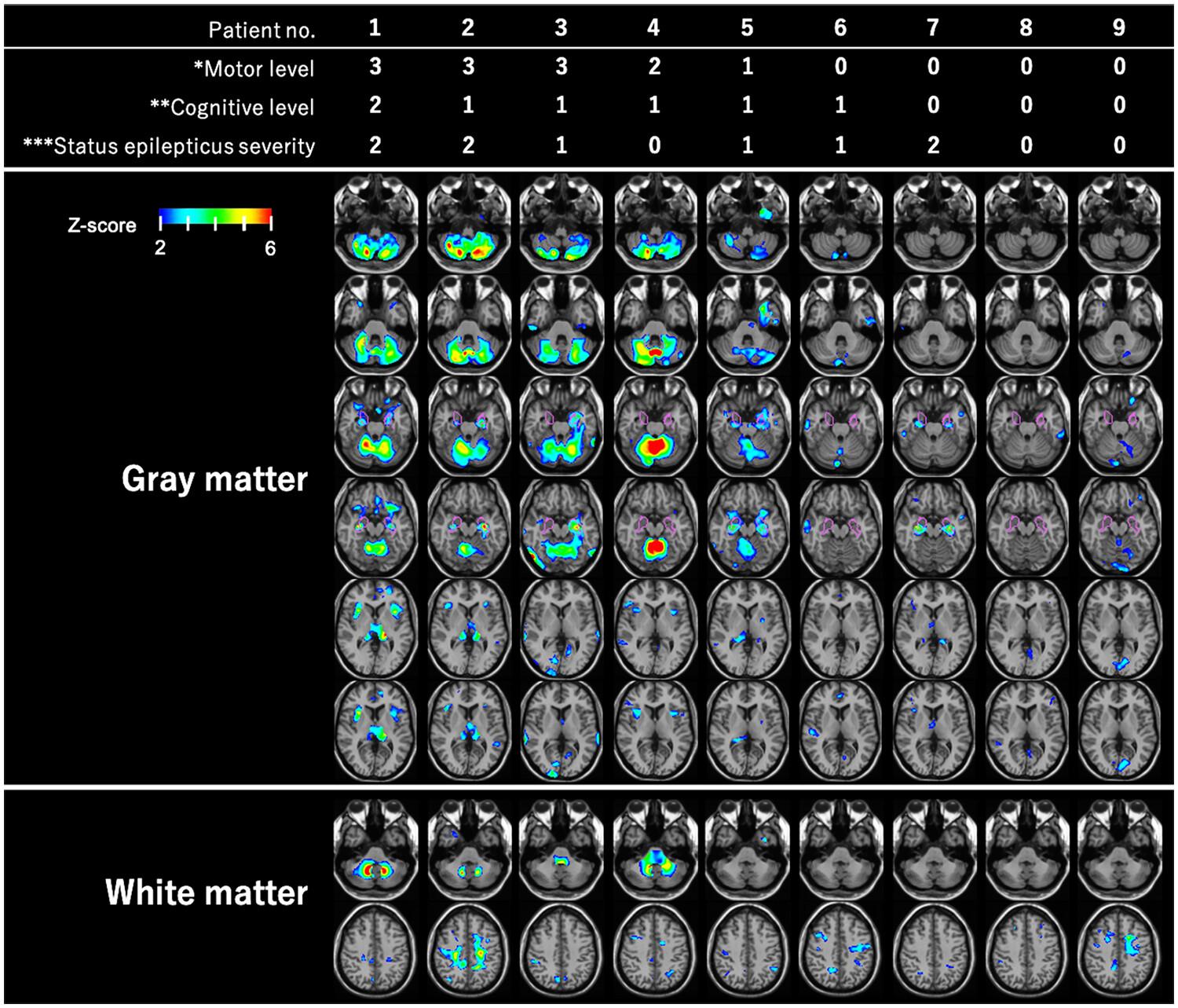
To investigate structural brain changes in gray matter (GM) and white matter (WM) volumes between 9 patients with AHC and 20 age-matched controls, VBM analysis was performed using three-dimensional T1-weighted magnetic resonance imaging. Single-case VBM analysis was also performed on nine patients with AHC to investigate the associations between the respective volumes of GM/WM differences and the motor level, cognitive level, and status epilepticus severity in patients with AHC.
Results
Compared with controls, patients with AHC showed significant GM volume reductions in both hippocampi and diffuse cerebellum, and there were WM reductions in both cerebral hemispheres. In patients with AHC, cases with more motor dysfunction, the less GM/WM volume of cerebellum was shown. Three of the six cases with cognitive dysfunction showed a clear GM volume reduction in the insulae. Five of the six cases with status epilepticus showed the GM volume reduction in hippocampi. One case had severe status epilepticus without motor dysfunction and showed no cerebellar atrophy.
Conclusion
With single-case VBM analysis, we could show the association between region-specific changes in brain volume and the severity of various clinical symptoms even in a small sample of subjects.
期刊介绍:
International Journal of Developmental Neuroscience publishes original research articles and critical review papers on all fundamental and clinical aspects of nervous system development, renewal and regeneration, as well as on the effects of genetic and environmental perturbations of brain development and homeostasis leading to neurodevelopmental disorders and neurological conditions. Studies describing the involvement of stem cells in nervous system maintenance and disease (including brain tumours), stem cell-based approaches for the investigation of neurodegenerative diseases, roles of neuroinflammation in development and disease, and neuroevolution are also encouraged. Investigations using molecular, cellular, physiological, genetic and epigenetic approaches in model systems ranging from simple invertebrates to human iPSC-based 2D and 3D models are encouraged, as are studies using experimental models that provide behavioural or evolutionary insights. The journal also publishes Special Issues dealing with topics at the cutting edge of research edited by Guest Editors appointed by the Editor in Chief. A major aim of the journal is to facilitate the transfer of fundamental studies of nervous system development, maintenance, and disease to clinical applications. The journal thus intends to disseminate valuable information for both biologists and physicians. International Journal of Developmental Neuroscience is owned and supported by The International Society for Developmental Neuroscience (ISDN), an organization of scientists interested in advancing developmental neuroscience research in the broadest sense.
 求助内容:
求助内容: 应助结果提醒方式:


