Gaye Keser, Ibrahim Sevki Bayrakdar, Filiz Namdar Pekiner, Özer Çelik, Kaan Orhan
{"title":"超声图像咬肌分割的深度学习方法。","authors":"Gaye Keser, Ibrahim Sevki Bayrakdar, Filiz Namdar Pekiner, Özer Çelik, Kaan Orhan","doi":"10.15557/jou.2022.0034","DOIUrl":null,"url":null,"abstract":"<p><strong>Aim: </strong>Deep learning algorithms have lately been used for medical image processing, and they have showed promise in a range of applications. The purpose of this study was to develop and test computer-based diagnostic tools for evaluating masseter muscle segmentation on ultrasonography images.</p><p><strong>Materials and methods: </strong>A total of 388 anonymous adult masseter muscle retrospective ultrasonographic images were evaluated. The masseter muscle was labeled on ultrasonography images using the polygonal type labeling method with the CranioCatch labeling program (CranioCatch, Eskişehir, Turkey). All images were re-checked and verified by Oral and Maxillofacial Radiology experts. This data set was divided into training (<i>n</i> = 312), verification (<i>n</i> = 38) and test (<i>n</i> = 38) sets. In the study, an artificial intelligence model was developed using PyTorch U-Net architecture, which is a deep learning approach.</p><p><strong>Results: </strong>In our study, the artificial intelligence deep learning model known as U-net provided the detection and segmentation of all test images, and when the success rate in the estimation of the images was evaluated, the F1, sensitivity and precision results of the model were 1.0, 1.0 and 1.0, respectively.</p><p><strong>Conclusion: </strong>Artificial intelligence shows promise in automatic segmentation of masseter muscle on ultrasonography images. This strategy can aid surgeons, radiologists, and other medical practitioners in reducing diagnostic time.</p>","PeriodicalId":45612,"journal":{"name":"Journal of Ultrasonography","volume":"22 91","pages":"e204-e208"},"PeriodicalIF":1.5000,"publicationDate":"2022-10-01","publicationTypes":"Journal Article","fieldsOfStudy":null,"isOpenAccess":false,"openAccessPdf":"https://ftp.ncbi.nlm.nih.gov/pub/pmc/oa_pdf/9a/25/jou-22-e204.PMC9714276.pdf","citationCount":"1","resultStr":"{\"title\":\"A Deep Learning Approach for Masseter Muscle Segmentation on Ultrasonography.\",\"authors\":\"Gaye Keser, Ibrahim Sevki Bayrakdar, Filiz Namdar Pekiner, Özer Çelik, Kaan Orhan\",\"doi\":\"10.15557/jou.2022.0034\",\"DOIUrl\":null,\"url\":null,\"abstract\":\"<p><strong>Aim: </strong>Deep learning algorithms have lately been used for medical image processing, and they have showed promise in a range of applications. The purpose of this study was to develop and test computer-based diagnostic tools for evaluating masseter muscle segmentation on ultrasonography images.</p><p><strong>Materials and methods: </strong>A total of 388 anonymous adult masseter muscle retrospective ultrasonographic images were evaluated. The masseter muscle was labeled on ultrasonography images using the polygonal type labeling method with the CranioCatch labeling program (CranioCatch, Eskişehir, Turkey). All images were re-checked and verified by Oral and Maxillofacial Radiology experts. This data set was divided into training (<i>n</i> = 312), verification (<i>n</i> = 38) and test (<i>n</i> = 38) sets. In the study, an artificial intelligence model was developed using PyTorch U-Net architecture, which is a deep learning approach.</p><p><strong>Results: </strong>In our study, the artificial intelligence deep learning model known as U-net provided the detection and segmentation of all test images, and when the success rate in the estimation of the images was evaluated, the F1, sensitivity and precision results of the model were 1.0, 1.0 and 1.0, respectively.</p><p><strong>Conclusion: </strong>Artificial intelligence shows promise in automatic segmentation of masseter muscle on ultrasonography images. This strategy can aid surgeons, radiologists, and other medical practitioners in reducing diagnostic time.</p>\",\"PeriodicalId\":45612,\"journal\":{\"name\":\"Journal of Ultrasonography\",\"volume\":\"22 91\",\"pages\":\"e204-e208\"},\"PeriodicalIF\":1.5000,\"publicationDate\":\"2022-10-01\",\"publicationTypes\":\"Journal Article\",\"fieldsOfStudy\":null,\"isOpenAccess\":false,\"openAccessPdf\":\"https://ftp.ncbi.nlm.nih.gov/pub/pmc/oa_pdf/9a/25/jou-22-e204.PMC9714276.pdf\",\"citationCount\":\"1\",\"resultStr\":null,\"platform\":\"Semanticscholar\",\"paperid\":null,\"PeriodicalName\":\"Journal of Ultrasonography\",\"FirstCategoryId\":\"1085\",\"ListUrlMain\":\"https://doi.org/10.15557/jou.2022.0034\",\"RegionNum\":0,\"RegionCategory\":null,\"ArticlePicture\":[],\"TitleCN\":null,\"AbstractTextCN\":null,\"PMCID\":null,\"EPubDate\":\"\",\"PubModel\":\"\",\"JCR\":\"Q3\",\"JCRName\":\"RADIOLOGY, NUCLEAR MEDICINE & MEDICAL IMAGING\",\"Score\":null,\"Total\":0}","platform":"Semanticscholar","paperid":null,"PeriodicalName":"Journal of Ultrasonography","FirstCategoryId":"1085","ListUrlMain":"https://doi.org/10.15557/jou.2022.0034","RegionNum":0,"RegionCategory":null,"ArticlePicture":[],"TitleCN":null,"AbstractTextCN":null,"PMCID":null,"EPubDate":"","PubModel":"","JCR":"Q3","JCRName":"RADIOLOGY, NUCLEAR MEDICINE & MEDICAL IMAGING","Score":null,"Total":0}
引用次数: 1
摘要
目的:深度学习算法最近被用于医学图像处理,并在一系列应用中显示出前景。本研究的目的是开发和测试基于计算机的诊断工具,以评估超声图像上的咬肌分割。材料与方法:对388张匿名成人咬肌回顾性超声图像进行评价。超声图像上的咬肌标记采用多边形标记法和CranioCatch标记程序(CranioCatch, eski ehir,土耳其)。所有图像由口腔颌面放射学专家重新检查和验证。该数据集分为训练集(n = 312)、验证集(n = 38)和测试集(n = 38)。在研究中,使用PyTorch U-Net架构开发了一个人工智能模型,这是一种深度学习方法。结果:在我们的研究中,人工智能深度学习模型U-net提供了所有测试图像的检测和分割,当评估图像估计的成功率时,模型的F1、灵敏度和精度结果分别为1.0、1.0和1.0。结论:人工智能在咬肌超声图像自动分割中具有广阔的应用前景。这一策略可以帮助外科医生、放射科医生和其他医疗从业者缩短诊断时间。
A Deep Learning Approach for Masseter Muscle Segmentation on Ultrasonography.
Aim: Deep learning algorithms have lately been used for medical image processing, and they have showed promise in a range of applications. The purpose of this study was to develop and test computer-based diagnostic tools for evaluating masseter muscle segmentation on ultrasonography images.
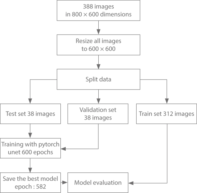
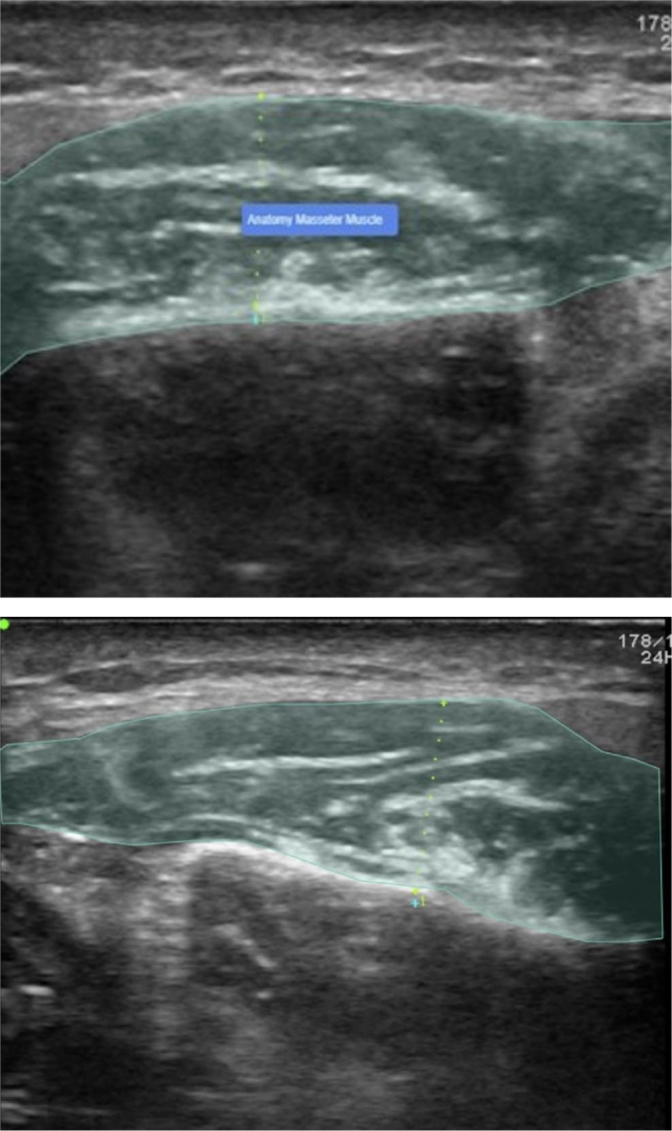
Materials and methods: A total of 388 anonymous adult masseter muscle retrospective ultrasonographic images were evaluated. The masseter muscle was labeled on ultrasonography images using the polygonal type labeling method with the CranioCatch labeling program (CranioCatch, Eskişehir, Turkey). All images were re-checked and verified by Oral and Maxillofacial Radiology experts. This data set was divided into training (n = 312), verification (n = 38) and test (n = 38) sets. In the study, an artificial intelligence model was developed using PyTorch U-Net architecture, which is a deep learning approach.
Results: In our study, the artificial intelligence deep learning model known as U-net provided the detection and segmentation of all test images, and when the success rate in the estimation of the images was evaluated, the F1, sensitivity and precision results of the model were 1.0, 1.0 and 1.0, respectively.
Conclusion: Artificial intelligence shows promise in automatic segmentation of masseter muscle on ultrasonography images. This strategy can aid surgeons, radiologists, and other medical practitioners in reducing diagnostic time.

 求助内容:
求助内容: 应助结果提醒方式:


