Dominique Segretain, Mathilde Di Marco, Chloé Dufeu, Diane Carette, Alain Trubuil, Georges Pointis
{"title":"协同细胞-细胞肌动蛋白网络重塑进行间隙连接内吞作用。","authors":"Dominique Segretain, Mathilde Di Marco, Chloé Dufeu, Diane Carette, Alain Trubuil, Georges Pointis","doi":"10.1186/s12610-023-00194-y","DOIUrl":null,"url":null,"abstract":"<p><strong>Background: </strong>The endocytosis of Gap junction plaques (GJP) requires cytoskeletal forces to internalize such large membranous structures. Actin, which partners the connexin proteins constituting Gap junctions and is located close to Annular Gap Junctions (AGJ), could be actively involved in this physiological process.</p><p><strong>Results: </strong>Electron Microscopy and Light Microscopy images, associated with time-lapse analysis and 3D reconstruction, used at high resolution and enhanced using ImageJ based software analysis, revealed that: i) actin cables, originating from Donor cells, insert on the edge of GJP and contribute to their invagination, giving rise to AGJ, whereas actin cables on the Acceptor cell side of the plaque are not modified; ii) actin cables from the Donor cell are continuous with the actin network present over the entire GJP surface. These actin cables fuse at a single point distant from the plaque, which then detaches itself from the membrane, condensing to form an actin mass during the final internalization process; iii) the Acceptor cell participates in the last step of the endocytic invagination process by forming an annular actin structure known as an actin ring.</p><p><strong>Conclusions: </strong>Together, these data suggest that the endocytosis of GJP is an example of a unique cooperative mechanism between the Donor (the traction of its actin cables) and the Acceptor cells (forming the actin ring).</p>","PeriodicalId":8730,"journal":{"name":"Basic and Clinical Andrology","volume":"33 1","pages":"20"},"PeriodicalIF":2.0000,"publicationDate":"2023-08-03","publicationTypes":"Journal Article","fieldsOfStudy":null,"isOpenAccess":false,"openAccessPdf":"https://www.ncbi.nlm.nih.gov/pmc/articles/PMC10399049/pdf/","citationCount":"0","resultStr":"{\"title\":\"Cooperative cell-cell actin network remodeling to perform Gap junction endocytosis.\",\"authors\":\"Dominique Segretain, Mathilde Di Marco, Chloé Dufeu, Diane Carette, Alain Trubuil, Georges Pointis\",\"doi\":\"10.1186/s12610-023-00194-y\",\"DOIUrl\":null,\"url\":null,\"abstract\":\"<p><strong>Background: </strong>The endocytosis of Gap junction plaques (GJP) requires cytoskeletal forces to internalize such large membranous structures. Actin, which partners the connexin proteins constituting Gap junctions and is located close to Annular Gap Junctions (AGJ), could be actively involved in this physiological process.</p><p><strong>Results: </strong>Electron Microscopy and Light Microscopy images, associated with time-lapse analysis and 3D reconstruction, used at high resolution and enhanced using ImageJ based software analysis, revealed that: i) actin cables, originating from Donor cells, insert on the edge of GJP and contribute to their invagination, giving rise to AGJ, whereas actin cables on the Acceptor cell side of the plaque are not modified; ii) actin cables from the Donor cell are continuous with the actin network present over the entire GJP surface. These actin cables fuse at a single point distant from the plaque, which then detaches itself from the membrane, condensing to form an actin mass during the final internalization process; iii) the Acceptor cell participates in the last step of the endocytic invagination process by forming an annular actin structure known as an actin ring.</p><p><strong>Conclusions: </strong>Together, these data suggest that the endocytosis of GJP is an example of a unique cooperative mechanism between the Donor (the traction of its actin cables) and the Acceptor cells (forming the actin ring).</p>\",\"PeriodicalId\":8730,\"journal\":{\"name\":\"Basic and Clinical Andrology\",\"volume\":\"33 1\",\"pages\":\"20\"},\"PeriodicalIF\":2.0000,\"publicationDate\":\"2023-08-03\",\"publicationTypes\":\"Journal Article\",\"fieldsOfStudy\":null,\"isOpenAccess\":false,\"openAccessPdf\":\"https://www.ncbi.nlm.nih.gov/pmc/articles/PMC10399049/pdf/\",\"citationCount\":\"0\",\"resultStr\":null,\"platform\":\"Semanticscholar\",\"paperid\":null,\"PeriodicalName\":\"Basic and Clinical Andrology\",\"FirstCategoryId\":\"3\",\"ListUrlMain\":\"https://doi.org/10.1186/s12610-023-00194-y\",\"RegionNum\":3,\"RegionCategory\":\"医学\",\"ArticlePicture\":[],\"TitleCN\":null,\"AbstractTextCN\":null,\"PMCID\":null,\"EPubDate\":\"\",\"PubModel\":\"\",\"JCR\":\"Q2\",\"JCRName\":\"ANDROLOGY\",\"Score\":null,\"Total\":0}","platform":"Semanticscholar","paperid":null,"PeriodicalName":"Basic and Clinical Andrology","FirstCategoryId":"3","ListUrlMain":"https://doi.org/10.1186/s12610-023-00194-y","RegionNum":3,"RegionCategory":"医学","ArticlePicture":[],"TitleCN":null,"AbstractTextCN":null,"PMCID":null,"EPubDate":"","PubModel":"","JCR":"Q2","JCRName":"ANDROLOGY","Score":null,"Total":0}
Cooperative cell-cell actin network remodeling to perform Gap junction endocytosis.
Background: The endocytosis of Gap junction plaques (GJP) requires cytoskeletal forces to internalize such large membranous structures. Actin, which partners the connexin proteins constituting Gap junctions and is located close to Annular Gap Junctions (AGJ), could be actively involved in this physiological process.
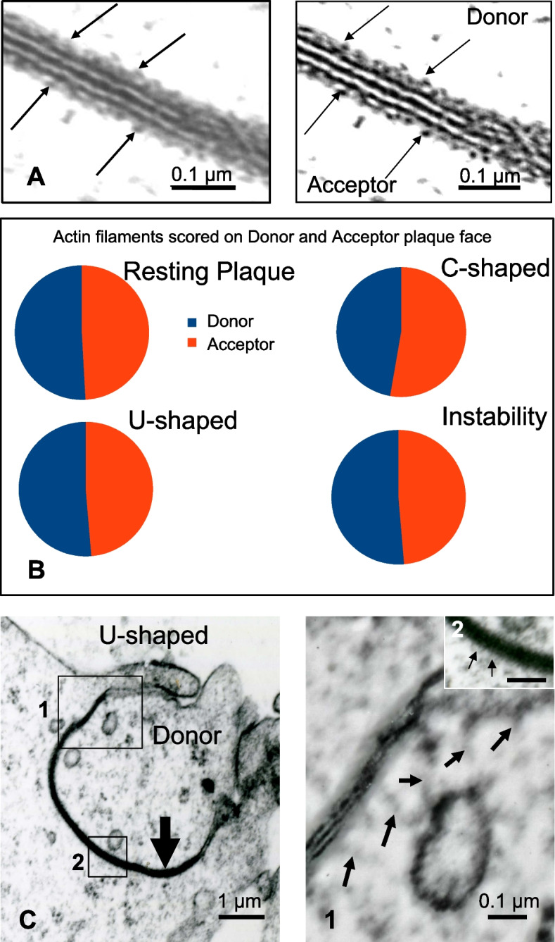
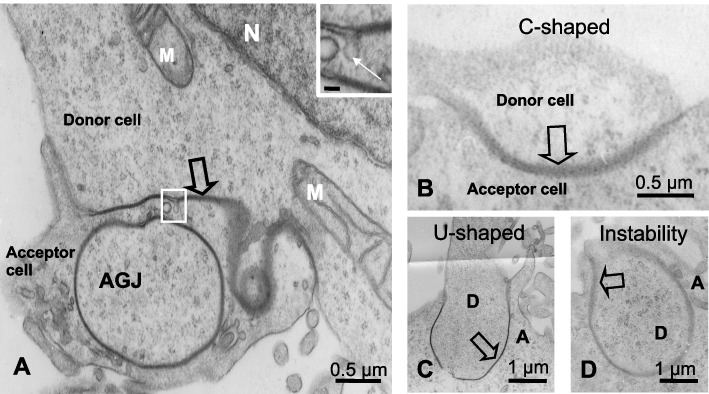
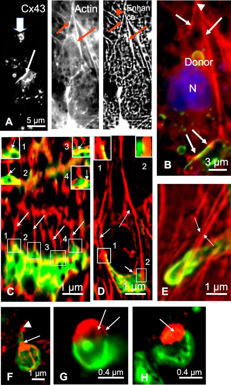
Results: Electron Microscopy and Light Microscopy images, associated with time-lapse analysis and 3D reconstruction, used at high resolution and enhanced using ImageJ based software analysis, revealed that: i) actin cables, originating from Donor cells, insert on the edge of GJP and contribute to their invagination, giving rise to AGJ, whereas actin cables on the Acceptor cell side of the plaque are not modified; ii) actin cables from the Donor cell are continuous with the actin network present over the entire GJP surface. These actin cables fuse at a single point distant from the plaque, which then detaches itself from the membrane, condensing to form an actin mass during the final internalization process; iii) the Acceptor cell participates in the last step of the endocytic invagination process by forming an annular actin structure known as an actin ring.
Conclusions: Together, these data suggest that the endocytosis of GJP is an example of a unique cooperative mechanism between the Donor (the traction of its actin cables) and the Acceptor cells (forming the actin ring).
期刊介绍:
Basic and Clinical Andrology is an open access journal in the domain of andrology covering all aspects of male reproductive and sexual health in both human and animal models. The journal aims to bring to light the various clinical advancements and research developments in andrology from the international community.

 求助内容:
求助内容: 应助结果提醒方式:


