Sini Toivonen, Miia Lehtinen, Peter Raivio, Juha Sinisalo, Antti Loimaala, Valtteri Uusitalo
{"title":"慢性冠状动脉疾病患者18F-FDG PET显示残留血管和脂肪组织炎症的存在","authors":"Sini Toivonen, Miia Lehtinen, Peter Raivio, Juha Sinisalo, Antti Loimaala, Valtteri Uusitalo","doi":"10.1007/s13139-022-00785-z","DOIUrl":null,"url":null,"abstract":"<p><strong>Purpose: </strong>We evaluated the residual vascular and adipose tissue inflammation in patients with chronic coronary artery disease (CAD) using positron emission tomography (PET).</p><p><strong>Methods: </strong>Our study population consisted of 98 patients with known CAD and 94 control subjects who had undergone <sup>18</sup>F-fluorodeoxyglucose (<sup>18</sup>F-FDG) PET due to non-cardiac reasons. Aortic root and vena cava superior <sup>18</sup>F-FDG uptake were measured to obtain the aortic root target-to-background ratio (TBR). In addition, adipose tissue PET measurements were done in pericoronary, epicardial, subcutaneous, and thoracic adipose tissue. Adipose tissue TBR was calculated using the left atrium as a reference region. Data are presented as mean ± standard deviation or as median (interquartile range).</p><p><strong>Results: </strong>The aortic root TBR was higher in CAD patients compared to control subjects, 1.68 (1.55-1.81) vs. 1.53 (1.43-1.64), <i>p</i> < 0.001. Subcutaneous adipose tissue uptake was elevated in CAD patients 0.30 (0.24-0.35) vs. 0.27 (0.23-0.31), <i>p</i> < 0.001. Metabolic activity of CAD patients and control subjects was comparable in the pericoronary (0.81 ± 0.18 vs. 0.80 ± 0.16, <i>p</i> = 0.59), epicardial (0.53 ± 0.21 vs. 0.51 ± 0.18, <i>p</i> = 0.38) and thoracic (0.31 ± 0.12 vs. 0.28 ± 0.12, <i>p</i> = 0.21) adipose tissue regions. Aortic root or adipose tissue <sup>18</sup>F-FDG uptake was not associated with the common CAD risk factors, coronary calcium score, or aortic calcium score (<i>p</i> value > 0.05).</p><p><strong>Conclusion: </strong>Patients with a chronic CAD had a higher aortic root and subcutaneous adipose tissue <sup>18</sup>F-FDG uptake compared to control patients, which suggests residual inflammatory risk.</p>","PeriodicalId":19384,"journal":{"name":"Nuclear Medicine and Molecular Imaging","volume":"57 3","pages":"117-125"},"PeriodicalIF":2.7000,"publicationDate":"2023-06-01","publicationTypes":"Journal Article","fieldsOfStudy":null,"isOpenAccess":false,"openAccessPdf":"https://www.ncbi.nlm.nih.gov/pmc/articles/PMC10172407/pdf/","citationCount":"0","resultStr":"{\"title\":\"The Presence of Residual Vascular and Adipose Tissue Inflammation on <sup>18</sup>F-FDG PET in Patients with Chronic Coronary Artery Disease.\",\"authors\":\"Sini Toivonen, Miia Lehtinen, Peter Raivio, Juha Sinisalo, Antti Loimaala, Valtteri Uusitalo\",\"doi\":\"10.1007/s13139-022-00785-z\",\"DOIUrl\":null,\"url\":null,\"abstract\":\"<p><strong>Purpose: </strong>We evaluated the residual vascular and adipose tissue inflammation in patients with chronic coronary artery disease (CAD) using positron emission tomography (PET).</p><p><strong>Methods: </strong>Our study population consisted of 98 patients with known CAD and 94 control subjects who had undergone <sup>18</sup>F-fluorodeoxyglucose (<sup>18</sup>F-FDG) PET due to non-cardiac reasons. Aortic root and vena cava superior <sup>18</sup>F-FDG uptake were measured to obtain the aortic root target-to-background ratio (TBR). In addition, adipose tissue PET measurements were done in pericoronary, epicardial, subcutaneous, and thoracic adipose tissue. Adipose tissue TBR was calculated using the left atrium as a reference region. Data are presented as mean ± standard deviation or as median (interquartile range).</p><p><strong>Results: </strong>The aortic root TBR was higher in CAD patients compared to control subjects, 1.68 (1.55-1.81) vs. 1.53 (1.43-1.64), <i>p</i> < 0.001. Subcutaneous adipose tissue uptake was elevated in CAD patients 0.30 (0.24-0.35) vs. 0.27 (0.23-0.31), <i>p</i> < 0.001. Metabolic activity of CAD patients and control subjects was comparable in the pericoronary (0.81 ± 0.18 vs. 0.80 ± 0.16, <i>p</i> = 0.59), epicardial (0.53 ± 0.21 vs. 0.51 ± 0.18, <i>p</i> = 0.38) and thoracic (0.31 ± 0.12 vs. 0.28 ± 0.12, <i>p</i> = 0.21) adipose tissue regions. Aortic root or adipose tissue <sup>18</sup>F-FDG uptake was not associated with the common CAD risk factors, coronary calcium score, or aortic calcium score (<i>p</i> value > 0.05).</p><p><strong>Conclusion: </strong>Patients with a chronic CAD had a higher aortic root and subcutaneous adipose tissue <sup>18</sup>F-FDG uptake compared to control patients, which suggests residual inflammatory risk.</p>\",\"PeriodicalId\":19384,\"journal\":{\"name\":\"Nuclear Medicine and Molecular Imaging\",\"volume\":\"57 3\",\"pages\":\"117-125\"},\"PeriodicalIF\":2.7000,\"publicationDate\":\"2023-06-01\",\"publicationTypes\":\"Journal Article\",\"fieldsOfStudy\":null,\"isOpenAccess\":false,\"openAccessPdf\":\"https://www.ncbi.nlm.nih.gov/pmc/articles/PMC10172407/pdf/\",\"citationCount\":\"0\",\"resultStr\":null,\"platform\":\"Semanticscholar\",\"paperid\":null,\"PeriodicalName\":\"Nuclear Medicine and Molecular Imaging\",\"FirstCategoryId\":\"1085\",\"ListUrlMain\":\"https://doi.org/10.1007/s13139-022-00785-z\",\"RegionNum\":0,\"RegionCategory\":null,\"ArticlePicture\":[],\"TitleCN\":null,\"AbstractTextCN\":null,\"PMCID\":null,\"EPubDate\":\"\",\"PubModel\":\"\",\"JCR\":\"Q3\",\"JCRName\":\"RADIOLOGY, NUCLEAR MEDICINE & MEDICAL IMAGING\",\"Score\":null,\"Total\":0}","platform":"Semanticscholar","paperid":null,"PeriodicalName":"Nuclear Medicine and Molecular Imaging","FirstCategoryId":"1085","ListUrlMain":"https://doi.org/10.1007/s13139-022-00785-z","RegionNum":0,"RegionCategory":null,"ArticlePicture":[],"TitleCN":null,"AbstractTextCN":null,"PMCID":null,"EPubDate":"","PubModel":"","JCR":"Q3","JCRName":"RADIOLOGY, NUCLEAR MEDICINE & MEDICAL IMAGING","Score":null,"Total":0}
The Presence of Residual Vascular and Adipose Tissue Inflammation on 18F-FDG PET in Patients with Chronic Coronary Artery Disease.
Purpose: We evaluated the residual vascular and adipose tissue inflammation in patients with chronic coronary artery disease (CAD) using positron emission tomography (PET).
Methods: Our study population consisted of 98 patients with known CAD and 94 control subjects who had undergone 18F-fluorodeoxyglucose (18F-FDG) PET due to non-cardiac reasons. Aortic root and vena cava superior 18F-FDG uptake were measured to obtain the aortic root target-to-background ratio (TBR). In addition, adipose tissue PET measurements were done in pericoronary, epicardial, subcutaneous, and thoracic adipose tissue. Adipose tissue TBR was calculated using the left atrium as a reference region. Data are presented as mean ± standard deviation or as median (interquartile range).
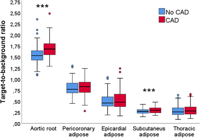
Results: The aortic root TBR was higher in CAD patients compared to control subjects, 1.68 (1.55-1.81) vs. 1.53 (1.43-1.64), p < 0.001. Subcutaneous adipose tissue uptake was elevated in CAD patients 0.30 (0.24-0.35) vs. 0.27 (0.23-0.31), p < 0.001. Metabolic activity of CAD patients and control subjects was comparable in the pericoronary (0.81 ± 0.18 vs. 0.80 ± 0.16, p = 0.59), epicardial (0.53 ± 0.21 vs. 0.51 ± 0.18, p = 0.38) and thoracic (0.31 ± 0.12 vs. 0.28 ± 0.12, p = 0.21) adipose tissue regions. Aortic root or adipose tissue 18F-FDG uptake was not associated with the common CAD risk factors, coronary calcium score, or aortic calcium score (p value > 0.05).
Conclusion: Patients with a chronic CAD had a higher aortic root and subcutaneous adipose tissue 18F-FDG uptake compared to control patients, which suggests residual inflammatory risk.
期刊介绍:
Nuclear Medicine and Molecular Imaging (Nucl Med Mol Imaging) is an official journal of the Korean Society of Nuclear Medicine, which bimonthly publishes papers on February, April, June, August, October, and December about nuclear medicine and related sciences such as radiochemistry, radiopharmacy, dosimetry and pharmacokinetics / pharmacodynamics of radiopharmaceuticals, nuclear and molecular imaging analysis, nuclear and molecular imaging instrumentation, radiation biology and radionuclide therapy. The journal specially welcomes works of artificial intelligence applied to nuclear medicine. The journal will also welcome original works relating to molecular imaging research such as the development of molecular imaging probes, reporter imaging assays, imaging cell trafficking, imaging endo(exo)genous gene expression, and imaging signal transduction. Nucl Med Mol Imaging publishes the following types of papers: original articles, reviews, case reports, editorials, interesting images, and letters to the editor.
The Korean Society of Nuclear Medicine (KSNM)
KSNM is a scientific and professional organization founded in 1961 and a member of the Korean Academy of Medical Sciences of the Korean Medical Association which was established by The Medical Services Law. The aims of KSNM are the promotion of nuclear medicine and cooperation of each member. The business of KSNM includes holding academic meetings and symposia, the publication of journals and books, planning and research of promoting science and health, and training and qualification of nuclear medicine specialists.
 求助内容:
求助内容: 应助结果提醒方式:


