Chie Ohnishi, Takashi Ohnishi, Peter Ntiamoah, Dara S. Ross, Masahiro Yamaguchi, Yukako Yagi
{"title":"标准化HER2免疫组织化学评估:校正染色和扫描引起的全片成像的颜色和强度变化","authors":"Chie Ohnishi, Takashi Ohnishi, Peter Ntiamoah, Dara S. Ross, Masahiro Yamaguchi, Yukako Yagi","doi":"10.1186/s42649-023-00091-8","DOIUrl":null,"url":null,"abstract":"<div><p>In the evaluation of human epidermal growth factor receptor 2 (HER2) immunohistochemistry (IHC) — one of the standard biomarkers for breast cancer— visual assessment is laborious and subjective. Image analysis using whole slide image (WSI) could produce more consistent results; however, color variability in WSIs due to the choice of stain and scanning processes may impact image analysis. We therefore developed a calibration protocol to diminish the staining and scanning variations of WSI using two calibrator slides. The IHC calibrator slide (IHC-CS) contains peptide-coated microbeads with different concentrations. The color distribution obtained from the WSI of stained IHC-CS reflects the staining process and scanner characteristics. A color chart slide (CCS) is also useful for calibrating the color variation due to the scanner. The results of the automated HER2 assessment were compared to confirm the effectiveness of two calibration slides. The IHC-CS and HER2 breast cancer cases were stained on different days. All stained slides and CCS were digitized by two different WSI scanners. Results revealed 100% concordance between automated evaluation and the pathologist’s assessment with both the scanner and staining calibration. The proposed method may enable consistent evaluation of HER2.</p></div>","PeriodicalId":470,"journal":{"name":"Applied Microscopy","volume":"53 1","pages":""},"PeriodicalIF":0.0000,"publicationDate":"2023-09-14","publicationTypes":"Journal Article","fieldsOfStudy":null,"isOpenAccess":false,"openAccessPdf":"https://www.ncbi.nlm.nih.gov/pmc/articles/PMC10499734/pdf/","citationCount":"0","resultStr":"{\"title\":\"Standardizing HER2 immunohistochemistry assessment: calibration of color and intensity variation in whole slide imaging caused by staining and scanning\",\"authors\":\"Chie Ohnishi, Takashi Ohnishi, Peter Ntiamoah, Dara S. Ross, Masahiro Yamaguchi, Yukako Yagi\",\"doi\":\"10.1186/s42649-023-00091-8\",\"DOIUrl\":null,\"url\":null,\"abstract\":\"<div><p>In the evaluation of human epidermal growth factor receptor 2 (HER2) immunohistochemistry (IHC) — one of the standard biomarkers for breast cancer— visual assessment is laborious and subjective. Image analysis using whole slide image (WSI) could produce more consistent results; however, color variability in WSIs due to the choice of stain and scanning processes may impact image analysis. We therefore developed a calibration protocol to diminish the staining and scanning variations of WSI using two calibrator slides. The IHC calibrator slide (IHC-CS) contains peptide-coated microbeads with different concentrations. The color distribution obtained from the WSI of stained IHC-CS reflects the staining process and scanner characteristics. A color chart slide (CCS) is also useful for calibrating the color variation due to the scanner. The results of the automated HER2 assessment were compared to confirm the effectiveness of two calibration slides. The IHC-CS and HER2 breast cancer cases were stained on different days. All stained slides and CCS were digitized by two different WSI scanners. Results revealed 100% concordance between automated evaluation and the pathologist’s assessment with both the scanner and staining calibration. The proposed method may enable consistent evaluation of HER2.</p></div>\",\"PeriodicalId\":470,\"journal\":{\"name\":\"Applied Microscopy\",\"volume\":\"53 1\",\"pages\":\"\"},\"PeriodicalIF\":0.0000,\"publicationDate\":\"2023-09-14\",\"publicationTypes\":\"Journal Article\",\"fieldsOfStudy\":null,\"isOpenAccess\":false,\"openAccessPdf\":\"https://www.ncbi.nlm.nih.gov/pmc/articles/PMC10499734/pdf/\",\"citationCount\":\"0\",\"resultStr\":null,\"platform\":\"Semanticscholar\",\"paperid\":null,\"PeriodicalName\":\"Applied Microscopy\",\"FirstCategoryId\":\"1085\",\"ListUrlMain\":\"https://link.springer.com/article/10.1186/s42649-023-00091-8\",\"RegionNum\":0,\"RegionCategory\":null,\"ArticlePicture\":[],\"TitleCN\":null,\"AbstractTextCN\":null,\"PMCID\":null,\"EPubDate\":\"\",\"PubModel\":\"\",\"JCR\":\"Q3\",\"JCRName\":\"Immunology and Microbiology\",\"Score\":null,\"Total\":0}","platform":"Semanticscholar","paperid":null,"PeriodicalName":"Applied Microscopy","FirstCategoryId":"1085","ListUrlMain":"https://link.springer.com/article/10.1186/s42649-023-00091-8","RegionNum":0,"RegionCategory":null,"ArticlePicture":[],"TitleCN":null,"AbstractTextCN":null,"PMCID":null,"EPubDate":"","PubModel":"","JCR":"Q3","JCRName":"Immunology and Microbiology","Score":null,"Total":0}
Standardizing HER2 immunohistochemistry assessment: calibration of color and intensity variation in whole slide imaging caused by staining and scanning
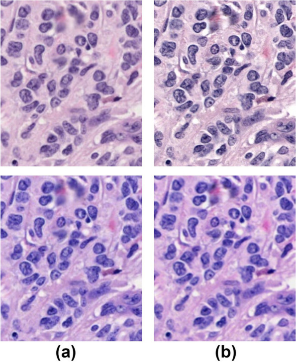
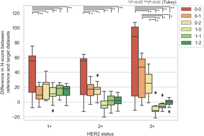
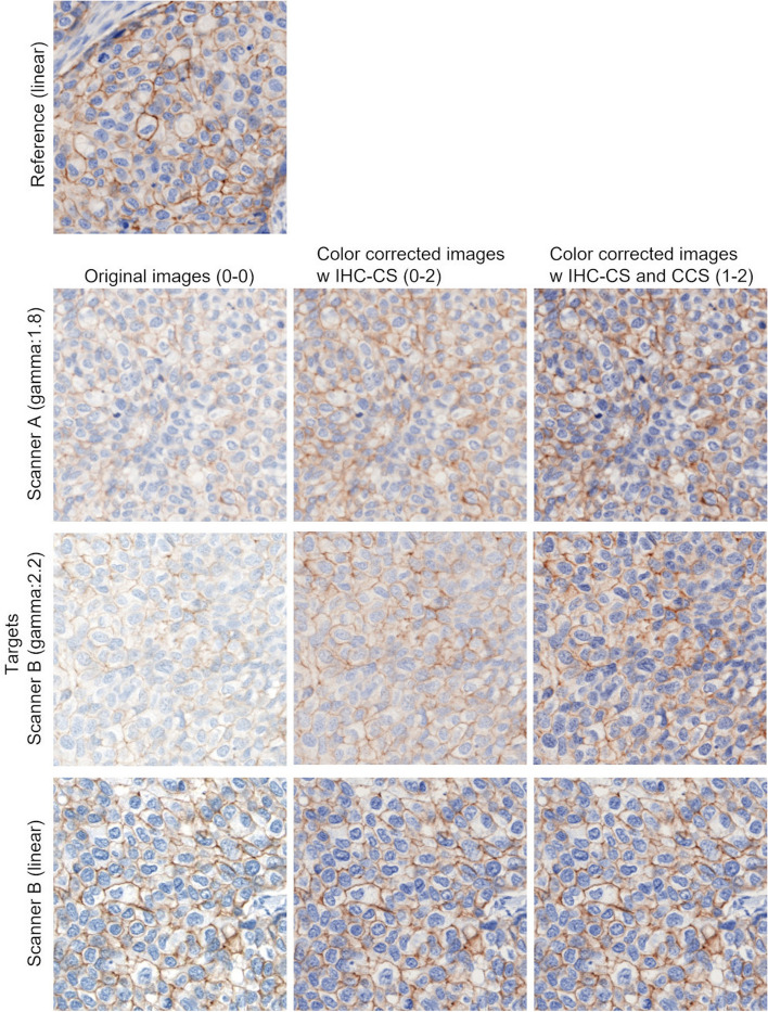
In the evaluation of human epidermal growth factor receptor 2 (HER2) immunohistochemistry (IHC) — one of the standard biomarkers for breast cancer— visual assessment is laborious and subjective. Image analysis using whole slide image (WSI) could produce more consistent results; however, color variability in WSIs due to the choice of stain and scanning processes may impact image analysis. We therefore developed a calibration protocol to diminish the staining and scanning variations of WSI using two calibrator slides. The IHC calibrator slide (IHC-CS) contains peptide-coated microbeads with different concentrations. The color distribution obtained from the WSI of stained IHC-CS reflects the staining process and scanner characteristics. A color chart slide (CCS) is also useful for calibrating the color variation due to the scanner. The results of the automated HER2 assessment were compared to confirm the effectiveness of two calibration slides. The IHC-CS and HER2 breast cancer cases were stained on different days. All stained slides and CCS were digitized by two different WSI scanners. Results revealed 100% concordance between automated evaluation and the pathologist’s assessment with both the scanner and staining calibration. The proposed method may enable consistent evaluation of HER2.
Applied MicroscopyImmunology and Microbiology-Applied Microbiology and Biotechnology
CiteScore
3.40
自引率
0.00%
发文量
10
审稿时长
10 weeks
期刊介绍:
Applied Microscopy is a peer-reviewed journal sponsored by the Korean Society of Microscopy. The journal covers all the interdisciplinary fields of technological developments in new microscopy methods and instrumentation and their applications to biological or materials science for determining structure and chemistry. ISSN: 22875123, 22874445.
 求助内容:
求助内容: 应助结果提醒方式:


