Vineet Gauhar, Daniele Castellani, Ben Hall Chew, Daron Smith, Chu Ann Chai, Khi Yung Fong, Jeremy Yuen-Chun Teoh, Olivier Traxer, Bhaskar Kumar Somani, Thomas Tailly
{"title":"未增强的计算机断层扫描作为逆行肾内手术后的成像标准是否矛盾地降低了结石的清除率并增加了残余碎片的额外治疗?TOWER组FLEXOR研究中5395例患者的结果。","authors":"Vineet Gauhar, Daniele Castellani, Ben Hall Chew, Daron Smith, Chu Ann Chai, Khi Yung Fong, Jeremy Yuen-Chun Teoh, Olivier Traxer, Bhaskar Kumar Somani, Thomas Tailly","doi":"10.1177/17562872231198629","DOIUrl":null,"url":null,"abstract":"<p><strong>Background: </strong>Assessment of residual fragments (RFs) is a key step after treatment of kidney stones.</p><p><strong>Objective: </strong>To evaluate differences in RFs estimation based on unenhanced computerized tomography (CT) <i>versus</i> X-rays/ultrasound after retrograde intrarenal surgery (RIRS) for kidney stones.</p><p><strong>Design: </strong>A retrospective analysis of data from 20 centers of adult patients who had RIRS was done (January 2018-August 2021).</p><p><strong>Methods: </strong>Exclusion criteria: ureteric stones, anomalous kidneys, bilateral renal stones. Patients were divided into two groups (group 1: CT; group 2: plain X-rays or combination of X-rays/ultrasound within 3 months after RIRS). Clinically significant RFs (CSRFs) were considered RFs ⩾ 4 mm. One-to-one propensity score matching for age, gender, and stone characteristics was performed. Multivariable logistic regression analysis was performed to evaluate independent predictors of CSRFs.</p><p><strong>Results: </strong>A total of 5395 patients were included (1748 in group 1; 3647 in group 2). After matching, 608 patients from each group with comparable baseline and stone characteristics were included. CSRFs were diagnosed in 1132 patients in the overall cohort (21.0%). Post-operative CT reported a significantly higher number of patients with RFs ⩾ 4 mm, before (35.7% <i>versus</i> 13.9%, <i>p</i> < 0.001) and after matching (43.1% <i>versus</i> 23.9%, <i>p</i> < 0.001). Only 21.8% of patients in the matched cohort had an ancillary procedure post-RIRS which was significantly higher in group 1 (74.8% <i>versus</i> 47.6%, <i>p</i> < 0.001). Age [OR 1.015 95% confidence interval (CI) 1.009-1.020, <i>p</i> < 0.001], stone size (OR 1.028 95% CI 1.017-1.040, <i>p</i> < 0.001), multiple stones (OR 1.171 95% CI 1.025-1.339, <i>p</i> = 0.021), lower pole stone (OR 1.853 95% CI 1.557-2.204, <i>p</i> < 0.001) and the use of post-operative CT scan (OR 5.9883 95% CI 5.094-7.037, <i>p</i> < 0.001) had significantly higher odds of having CSRFs.</p><p><strong>Conclusions: </strong>CT is the only reliable imaging to assess the burden of RFs following RIRS and urologist should consider at least one CT scan to determine the same and definitely plan reintervention only based on CT rather than ultrasound and X-ray combination.</p>","PeriodicalId":23010,"journal":{"name":"Therapeutic Advances in Urology","volume":"15 ","pages":"17562872231198629"},"PeriodicalIF":2.6000,"publicationDate":"2023-01-01","publicationTypes":"Journal Article","fieldsOfStudy":null,"isOpenAccess":false,"openAccessPdf":"https://ftp.ncbi.nlm.nih.gov/pub/pmc/oa_pdf/fd/ce/10.1177_17562872231198629.PMC10493056.pdf","citationCount":"0","resultStr":"{\"title\":\"Does unenhanced computerized tomography as imaging standard post-retrograde intrarenal surgery paradoxically reduce stone-free rate and increase additional treatment for residual fragments? Outcomes from 5395 patients in the FLEXOR study by the TOWER group.\",\"authors\":\"Vineet Gauhar, Daniele Castellani, Ben Hall Chew, Daron Smith, Chu Ann Chai, Khi Yung Fong, Jeremy Yuen-Chun Teoh, Olivier Traxer, Bhaskar Kumar Somani, Thomas Tailly\",\"doi\":\"10.1177/17562872231198629\",\"DOIUrl\":null,\"url\":null,\"abstract\":\"<p><strong>Background: </strong>Assessment of residual fragments (RFs) is a key step after treatment of kidney stones.</p><p><strong>Objective: </strong>To evaluate differences in RFs estimation based on unenhanced computerized tomography (CT) <i>versus</i> X-rays/ultrasound after retrograde intrarenal surgery (RIRS) for kidney stones.</p><p><strong>Design: </strong>A retrospective analysis of data from 20 centers of adult patients who had RIRS was done (January 2018-August 2021).</p><p><strong>Methods: </strong>Exclusion criteria: ureteric stones, anomalous kidneys, bilateral renal stones. Patients were divided into two groups (group 1: CT; group 2: plain X-rays or combination of X-rays/ultrasound within 3 months after RIRS). Clinically significant RFs (CSRFs) were considered RFs ⩾ 4 mm. One-to-one propensity score matching for age, gender, and stone characteristics was performed. Multivariable logistic regression analysis was performed to evaluate independent predictors of CSRFs.</p><p><strong>Results: </strong>A total of 5395 patients were included (1748 in group 1; 3647 in group 2). After matching, 608 patients from each group with comparable baseline and stone characteristics were included. CSRFs were diagnosed in 1132 patients in the overall cohort (21.0%). Post-operative CT reported a significantly higher number of patients with RFs ⩾ 4 mm, before (35.7% <i>versus</i> 13.9%, <i>p</i> < 0.001) and after matching (43.1% <i>versus</i> 23.9%, <i>p</i> < 0.001). Only 21.8% of patients in the matched cohort had an ancillary procedure post-RIRS which was significantly higher in group 1 (74.8% <i>versus</i> 47.6%, <i>p</i> < 0.001). Age [OR 1.015 95% confidence interval (CI) 1.009-1.020, <i>p</i> < 0.001], stone size (OR 1.028 95% CI 1.017-1.040, <i>p</i> < 0.001), multiple stones (OR 1.171 95% CI 1.025-1.339, <i>p</i> = 0.021), lower pole stone (OR 1.853 95% CI 1.557-2.204, <i>p</i> < 0.001) and the use of post-operative CT scan (OR 5.9883 95% CI 5.094-7.037, <i>p</i> < 0.001) had significantly higher odds of having CSRFs.</p><p><strong>Conclusions: </strong>CT is the only reliable imaging to assess the burden of RFs following RIRS and urologist should consider at least one CT scan to determine the same and definitely plan reintervention only based on CT rather than ultrasound and X-ray combination.</p>\",\"PeriodicalId\":23010,\"journal\":{\"name\":\"Therapeutic Advances in Urology\",\"volume\":\"15 \",\"pages\":\"17562872231198629\"},\"PeriodicalIF\":2.6000,\"publicationDate\":\"2023-01-01\",\"publicationTypes\":\"Journal Article\",\"fieldsOfStudy\":null,\"isOpenAccess\":false,\"openAccessPdf\":\"https://ftp.ncbi.nlm.nih.gov/pub/pmc/oa_pdf/fd/ce/10.1177_17562872231198629.PMC10493056.pdf\",\"citationCount\":\"0\",\"resultStr\":null,\"platform\":\"Semanticscholar\",\"paperid\":null,\"PeriodicalName\":\"Therapeutic Advances in Urology\",\"FirstCategoryId\":\"3\",\"ListUrlMain\":\"https://doi.org/10.1177/17562872231198629\",\"RegionNum\":4,\"RegionCategory\":\"医学\",\"ArticlePicture\":[],\"TitleCN\":null,\"AbstractTextCN\":null,\"PMCID\":null,\"EPubDate\":\"\",\"PubModel\":\"\",\"JCR\":\"Q2\",\"JCRName\":\"UROLOGY & NEPHROLOGY\",\"Score\":null,\"Total\":0}","platform":"Semanticscholar","paperid":null,"PeriodicalName":"Therapeutic Advances in Urology","FirstCategoryId":"3","ListUrlMain":"https://doi.org/10.1177/17562872231198629","RegionNum":4,"RegionCategory":"医学","ArticlePicture":[],"TitleCN":null,"AbstractTextCN":null,"PMCID":null,"EPubDate":"","PubModel":"","JCR":"Q2","JCRName":"UROLOGY & NEPHROLOGY","Score":null,"Total":0}
引用次数: 0
摘要
背景:残留碎片(RFs)的评估是肾结石治疗后的关键步骤。目的:评价肾结石逆行肾内手术(RIRS)后基于非增强计算机断层扫描(CT)与x射线/超声的RFs估计的差异。设计:回顾性分析来自20个中心的RIRS成年患者的数据(2018年1月至2021年8月)。方法:排除标准:输尿管结石、肾异常、双侧肾结石。患者分为两组(1组:CT;第二组:术后3个月内进行x光平片或x光/超声联合检查)。临床显著的RFs (CSRFs)被认为是小于4 mm的RFs。对年龄、性别和结石特征进行一对一的倾向评分匹配。采用多变量logistic回归分析评价csrf的独立预测因子。结果:共纳入5395例患者(1组1748例;2组3647例)。匹配后,每组608例基线和结石特征相似的患者入组。在整个队列中,1132例(21.0%)患者被诊断为csrf。患者术后CT报告数量明显高于RFs⩾4毫米,之前(35.7%比13.9%,p与23.9%,p与47.6%,p p p p = 0.021),低杆石(或1.853 95%可信区间1.557 - -2.204,p p结论:CT是唯一可靠的成像评估后RFs rir的负担和泌尿科医生应该考虑至少一个CT扫描来确定相同的和绝对计划reintervention只基于CT而非超声和x射线组合。
Does unenhanced computerized tomography as imaging standard post-retrograde intrarenal surgery paradoxically reduce stone-free rate and increase additional treatment for residual fragments? Outcomes from 5395 patients in the FLEXOR study by the TOWER group.
Background: Assessment of residual fragments (RFs) is a key step after treatment of kidney stones.
Objective: To evaluate differences in RFs estimation based on unenhanced computerized tomography (CT) versus X-rays/ultrasound after retrograde intrarenal surgery (RIRS) for kidney stones.
Design: A retrospective analysis of data from 20 centers of adult patients who had RIRS was done (January 2018-August 2021).
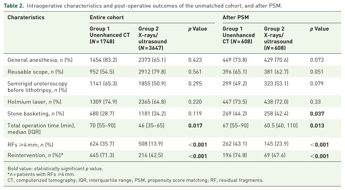
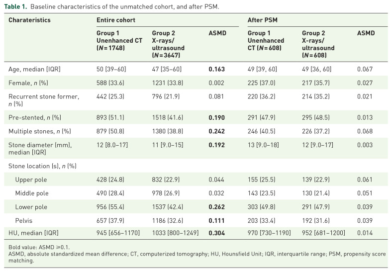
Methods: Exclusion criteria: ureteric stones, anomalous kidneys, bilateral renal stones. Patients were divided into two groups (group 1: CT; group 2: plain X-rays or combination of X-rays/ultrasound within 3 months after RIRS). Clinically significant RFs (CSRFs) were considered RFs ⩾ 4 mm. One-to-one propensity score matching for age, gender, and stone characteristics was performed. Multivariable logistic regression analysis was performed to evaluate independent predictors of CSRFs.
Results: A total of 5395 patients were included (1748 in group 1; 3647 in group 2). After matching, 608 patients from each group with comparable baseline and stone characteristics were included. CSRFs were diagnosed in 1132 patients in the overall cohort (21.0%). Post-operative CT reported a significantly higher number of patients with RFs ⩾ 4 mm, before (35.7% versus 13.9%, p < 0.001) and after matching (43.1% versus 23.9%, p < 0.001). Only 21.8% of patients in the matched cohort had an ancillary procedure post-RIRS which was significantly higher in group 1 (74.8% versus 47.6%, p < 0.001). Age [OR 1.015 95% confidence interval (CI) 1.009-1.020, p < 0.001], stone size (OR 1.028 95% CI 1.017-1.040, p < 0.001), multiple stones (OR 1.171 95% CI 1.025-1.339, p = 0.021), lower pole stone (OR 1.853 95% CI 1.557-2.204, p < 0.001) and the use of post-operative CT scan (OR 5.9883 95% CI 5.094-7.037, p < 0.001) had significantly higher odds of having CSRFs.
Conclusions: CT is the only reliable imaging to assess the burden of RFs following RIRS and urologist should consider at least one CT scan to determine the same and definitely plan reintervention only based on CT rather than ultrasound and X-ray combination.
期刊介绍:
Therapeutic Advances in Urology delivers the highest quality peer-reviewed articles, reviews, and scholarly comment on pioneering efforts and innovative studies across all areas of urology.
The journal has a strong clinical and pharmacological focus and is aimed at clinicians and researchers in urology, providing a forum in print and online for publishing the highest quality articles in this area. The editors welcome articles of current interest across all areas of urology, including treatment of urological disorders, with a focus on emerging pharmacological therapies.

 求助内容:
求助内容: 应助结果提醒方式:


