Magdalena Posadzy, Monika Ostrowska, Emil Michalski, Piotr Gietka, Małgorzata Mańczak, Michał Lanckoroński, Marek Leszkiewicz, Iwona Sudoł-Szopińska
{"title":"新诊断为幼年特发性关节炎的儿童和青少年足部超声和MRI。","authors":"Magdalena Posadzy, Monika Ostrowska, Emil Michalski, Piotr Gietka, Małgorzata Mańczak, Michał Lanckoroński, Marek Leszkiewicz, Iwona Sudoł-Szopińska","doi":"10.15557/jou.2023.0019","DOIUrl":null,"url":null,"abstract":"<p><strong>Aim: </strong>To evaluate the spectrum of inflammatory features in foot joints which may be detected on routinely performed ultrasound (US) and magnetic resonance imaging (MRI) in children newly diagnosed with juvenile idiopathic arthritis (JIA).</p><p><strong>Material and methods: </strong>Two groups of children hospitalized in a reference center for rheumatology, newly diagnosed with JIA and suspected of foot involvement in the course of JIA were included in this retrospective study. In the first group of 47 patients aged 1-18 years, the imaging was restricted to US. The second group of 22 patients aged 5-18 years underwent only non-contrast MRI of the foot.</p><p><strong>Results: </strong>The most frequent pathologies seen on US included effusion and synovial thickening in the first metatarsophalangeal joint (MTP1), followed by the tibiotalar joint. Synovial hyperemia on color Doppler US images was present most frequently in the Chopart and midtarsal joints (64%; 7/11 cases), followed by the tibiotalar joint (45%; 5/11), and MTP2-5 joint synovitis (40%; 4/10). Grade 3 hyperemia was present only in four cases; grades 1 and 2 were detected in the majority of cases. On MRI, bone marrow edema was the most frequent pathology, found mostly in the calcaneus (45%; 10/22 cases), while alterations of the forefoot were rare. No cases of bursitis, enthesitis, cysts, erosions or ankylosis were diagnosed in either of the analyzed groups.</p><p><strong>Conclusions: </strong>Routine US of the foot is recommended for early detection of its involvement in JIA in daily clinical practice. Although MRI can identify features of various JIA stages, it is particularly useful for the detection of bone marrow alterations.</p>","PeriodicalId":45612,"journal":{"name":"Journal of Ultrasonography","volume":"23 94","pages":"e106-e113"},"PeriodicalIF":1.5000,"publicationDate":"2023-09-01","publicationTypes":"Journal Article","fieldsOfStudy":null,"isOpenAccess":false,"openAccessPdf":"https://ftp.ncbi.nlm.nih.gov/pub/pmc/oa_pdf/1f/96/jou-23-94-jou.2023.0019.PMC10494807.pdf","citationCount":"0","resultStr":"{\"title\":\"Ultrasound and MRI of the foot in children and adolescents newly diagnosed with juvenile idiopathic arthritis.\",\"authors\":\"Magdalena Posadzy, Monika Ostrowska, Emil Michalski, Piotr Gietka, Małgorzata Mańczak, Michał Lanckoroński, Marek Leszkiewicz, Iwona Sudoł-Szopińska\",\"doi\":\"10.15557/jou.2023.0019\",\"DOIUrl\":null,\"url\":null,\"abstract\":\"<p><strong>Aim: </strong>To evaluate the spectrum of inflammatory features in foot joints which may be detected on routinely performed ultrasound (US) and magnetic resonance imaging (MRI) in children newly diagnosed with juvenile idiopathic arthritis (JIA).</p><p><strong>Material and methods: </strong>Two groups of children hospitalized in a reference center for rheumatology, newly diagnosed with JIA and suspected of foot involvement in the course of JIA were included in this retrospective study. In the first group of 47 patients aged 1-18 years, the imaging was restricted to US. The second group of 22 patients aged 5-18 years underwent only non-contrast MRI of the foot.</p><p><strong>Results: </strong>The most frequent pathologies seen on US included effusion and synovial thickening in the first metatarsophalangeal joint (MTP1), followed by the tibiotalar joint. Synovial hyperemia on color Doppler US images was present most frequently in the Chopart and midtarsal joints (64%; 7/11 cases), followed by the tibiotalar joint (45%; 5/11), and MTP2-5 joint synovitis (40%; 4/10). Grade 3 hyperemia was present only in four cases; grades 1 and 2 were detected in the majority of cases. On MRI, bone marrow edema was the most frequent pathology, found mostly in the calcaneus (45%; 10/22 cases), while alterations of the forefoot were rare. No cases of bursitis, enthesitis, cysts, erosions or ankylosis were diagnosed in either of the analyzed groups.</p><p><strong>Conclusions: </strong>Routine US of the foot is recommended for early detection of its involvement in JIA in daily clinical practice. Although MRI can identify features of various JIA stages, it is particularly useful for the detection of bone marrow alterations.</p>\",\"PeriodicalId\":45612,\"journal\":{\"name\":\"Journal of Ultrasonography\",\"volume\":\"23 94\",\"pages\":\"e106-e113\"},\"PeriodicalIF\":1.5000,\"publicationDate\":\"2023-09-01\",\"publicationTypes\":\"Journal Article\",\"fieldsOfStudy\":null,\"isOpenAccess\":false,\"openAccessPdf\":\"https://ftp.ncbi.nlm.nih.gov/pub/pmc/oa_pdf/1f/96/jou-23-94-jou.2023.0019.PMC10494807.pdf\",\"citationCount\":\"0\",\"resultStr\":null,\"platform\":\"Semanticscholar\",\"paperid\":null,\"PeriodicalName\":\"Journal of Ultrasonography\",\"FirstCategoryId\":\"1085\",\"ListUrlMain\":\"https://doi.org/10.15557/jou.2023.0019\",\"RegionNum\":0,\"RegionCategory\":null,\"ArticlePicture\":[],\"TitleCN\":null,\"AbstractTextCN\":null,\"PMCID\":null,\"EPubDate\":\"\",\"PubModel\":\"\",\"JCR\":\"Q3\",\"JCRName\":\"RADIOLOGY, NUCLEAR MEDICINE & MEDICAL IMAGING\",\"Score\":null,\"Total\":0}","platform":"Semanticscholar","paperid":null,"PeriodicalName":"Journal of Ultrasonography","FirstCategoryId":"1085","ListUrlMain":"https://doi.org/10.15557/jou.2023.0019","RegionNum":0,"RegionCategory":null,"ArticlePicture":[],"TitleCN":null,"AbstractTextCN":null,"PMCID":null,"EPubDate":"","PubModel":"","JCR":"Q3","JCRName":"RADIOLOGY, NUCLEAR MEDICINE & MEDICAL IMAGING","Score":null,"Total":0}
Ultrasound and MRI of the foot in children and adolescents newly diagnosed with juvenile idiopathic arthritis.
Aim: To evaluate the spectrum of inflammatory features in foot joints which may be detected on routinely performed ultrasound (US) and magnetic resonance imaging (MRI) in children newly diagnosed with juvenile idiopathic arthritis (JIA).
Material and methods: Two groups of children hospitalized in a reference center for rheumatology, newly diagnosed with JIA and suspected of foot involvement in the course of JIA were included in this retrospective study. In the first group of 47 patients aged 1-18 years, the imaging was restricted to US. The second group of 22 patients aged 5-18 years underwent only non-contrast MRI of the foot.
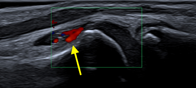
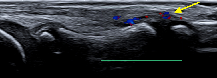
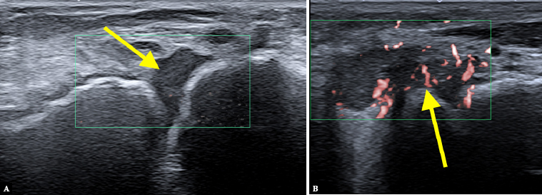
Results: The most frequent pathologies seen on US included effusion and synovial thickening in the first metatarsophalangeal joint (MTP1), followed by the tibiotalar joint. Synovial hyperemia on color Doppler US images was present most frequently in the Chopart and midtarsal joints (64%; 7/11 cases), followed by the tibiotalar joint (45%; 5/11), and MTP2-5 joint synovitis (40%; 4/10). Grade 3 hyperemia was present only in four cases; grades 1 and 2 were detected in the majority of cases. On MRI, bone marrow edema was the most frequent pathology, found mostly in the calcaneus (45%; 10/22 cases), while alterations of the forefoot were rare. No cases of bursitis, enthesitis, cysts, erosions or ankylosis were diagnosed in either of the analyzed groups.
Conclusions: Routine US of the foot is recommended for early detection of its involvement in JIA in daily clinical practice. Although MRI can identify features of various JIA stages, it is particularly useful for the detection of bone marrow alterations.

 求助内容:
求助内容: 应助结果提醒方式:


