Ramesh Venkatesh, Ashit Handa, Vishma Prabhu, Sai Prashanti Chitturi, Aishwarya Joshi, Isha Acharya, Rubble Mangla, Naresh Kumar Yadav, Jay Chhablani
{"title":"中央后透明体纤维化:演变和结果。","authors":"Ramesh Venkatesh, Ashit Handa, Vishma Prabhu, Sai Prashanti Chitturi, Aishwarya Joshi, Isha Acharya, Rubble Mangla, Naresh Kumar Yadav, Jay Chhablani","doi":"10.1186/s40942-023-00494-5","DOIUrl":null,"url":null,"abstract":"<p><strong>Purpose: </strong>To report contributory factors and clinical outcomes of central posterior hyaloid fibrosis (CPHF) associated with neovascular age-related macular degeneration (nAMD).</p><p><strong>Methods: </strong>In this retrospective, single-center study, patients with CPHF and nAMD were included. Demographic and imaging characteristics, as well as the anatomical and functional outcomes, of these patients were analysed.</p><p><strong>Results: </strong>We identified 530 eyes in 273 patients with chronic predominantly scarred macular neovascularisation (MNV), and 32 eyes in 29 patients revealed CPHF, representing a prevalence of 6%. Patients had a mean age of 72.76 years. Before and during the development of CPHF, Type 2 MNV was observed in all eyes. At the time of MNV diagnosis, mean logMAR visual acuity was 1.308 ± 0.559 (20/407). The average time to develop CPHF was 27.3 months since the diagnosis of MNV. At the time of CPHF identification, the mean logMAR visual acuity was 1.498 ± 0.374 (20/630). RPE tear was observed in 6% (n = 2) of CPHF eyes. The average number of intravitreal anti-VEGF injections administered prior to the diagnosis of CPHF was 2.4 and after the onset of CPHF was 0.9. The final visual acuity was not significantly different at the final follow-up visit [1.304 ± 0.42 (20/402); p = 0.646].</p><p><strong>Conclusion: </strong>Rarely observed in eyes with predominantly scarred subfoveal type 2 MNVs and extensive subretinal fibrosis, CPHF is associated with poor visual outcomes. Its presence could possibly suggest a profibrotic effect of MNV on the posterior hyaloid.</p><p><strong>Trial registration number: </strong>Not applicable.</p>","PeriodicalId":14289,"journal":{"name":"International Journal of Retina and Vitreous","volume":null,"pages":null},"PeriodicalIF":1.9000,"publicationDate":"2023-09-07","publicationTypes":"Journal Article","fieldsOfStudy":null,"isOpenAccess":false,"openAccessPdf":"https://www.ncbi.nlm.nih.gov/pmc/articles/PMC10486079/pdf/","citationCount":"0","resultStr":"{\"title\":\"Central posterior hyaloid fibrosis: evolution and outcomes.\",\"authors\":\"Ramesh Venkatesh, Ashit Handa, Vishma Prabhu, Sai Prashanti Chitturi, Aishwarya Joshi, Isha Acharya, Rubble Mangla, Naresh Kumar Yadav, Jay Chhablani\",\"doi\":\"10.1186/s40942-023-00494-5\",\"DOIUrl\":null,\"url\":null,\"abstract\":\"<p><strong>Purpose: </strong>To report contributory factors and clinical outcomes of central posterior hyaloid fibrosis (CPHF) associated with neovascular age-related macular degeneration (nAMD).</p><p><strong>Methods: </strong>In this retrospective, single-center study, patients with CPHF and nAMD were included. Demographic and imaging characteristics, as well as the anatomical and functional outcomes, of these patients were analysed.</p><p><strong>Results: </strong>We identified 530 eyes in 273 patients with chronic predominantly scarred macular neovascularisation (MNV), and 32 eyes in 29 patients revealed CPHF, representing a prevalence of 6%. Patients had a mean age of 72.76 years. Before and during the development of CPHF, Type 2 MNV was observed in all eyes. At the time of MNV diagnosis, mean logMAR visual acuity was 1.308 ± 0.559 (20/407). The average time to develop CPHF was 27.3 months since the diagnosis of MNV. At the time of CPHF identification, the mean logMAR visual acuity was 1.498 ± 0.374 (20/630). RPE tear was observed in 6% (n = 2) of CPHF eyes. The average number of intravitreal anti-VEGF injections administered prior to the diagnosis of CPHF was 2.4 and after the onset of CPHF was 0.9. The final visual acuity was not significantly different at the final follow-up visit [1.304 ± 0.42 (20/402); p = 0.646].</p><p><strong>Conclusion: </strong>Rarely observed in eyes with predominantly scarred subfoveal type 2 MNVs and extensive subretinal fibrosis, CPHF is associated with poor visual outcomes. Its presence could possibly suggest a profibrotic effect of MNV on the posterior hyaloid.</p><p><strong>Trial registration number: </strong>Not applicable.</p>\",\"PeriodicalId\":14289,\"journal\":{\"name\":\"International Journal of Retina and Vitreous\",\"volume\":null,\"pages\":null},\"PeriodicalIF\":1.9000,\"publicationDate\":\"2023-09-07\",\"publicationTypes\":\"Journal Article\",\"fieldsOfStudy\":null,\"isOpenAccess\":false,\"openAccessPdf\":\"https://www.ncbi.nlm.nih.gov/pmc/articles/PMC10486079/pdf/\",\"citationCount\":\"0\",\"resultStr\":null,\"platform\":\"Semanticscholar\",\"paperid\":null,\"PeriodicalName\":\"International Journal of Retina and Vitreous\",\"FirstCategoryId\":\"1085\",\"ListUrlMain\":\"https://doi.org/10.1186/s40942-023-00494-5\",\"RegionNum\":0,\"RegionCategory\":null,\"ArticlePicture\":[],\"TitleCN\":null,\"AbstractTextCN\":null,\"PMCID\":null,\"EPubDate\":\"\",\"PubModel\":\"\",\"JCR\":\"Q2\",\"JCRName\":\"OPHTHALMOLOGY\",\"Score\":null,\"Total\":0}","platform":"Semanticscholar","paperid":null,"PeriodicalName":"International Journal of Retina and Vitreous","FirstCategoryId":"1085","ListUrlMain":"https://doi.org/10.1186/s40942-023-00494-5","RegionNum":0,"RegionCategory":null,"ArticlePicture":[],"TitleCN":null,"AbstractTextCN":null,"PMCID":null,"EPubDate":"","PubModel":"","JCR":"Q2","JCRName":"OPHTHALMOLOGY","Score":null,"Total":0}
Central posterior hyaloid fibrosis: evolution and outcomes.
Purpose: To report contributory factors and clinical outcomes of central posterior hyaloid fibrosis (CPHF) associated with neovascular age-related macular degeneration (nAMD).
Methods: In this retrospective, single-center study, patients with CPHF and nAMD were included. Demographic and imaging characteristics, as well as the anatomical and functional outcomes, of these patients were analysed.
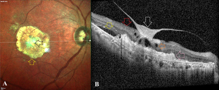
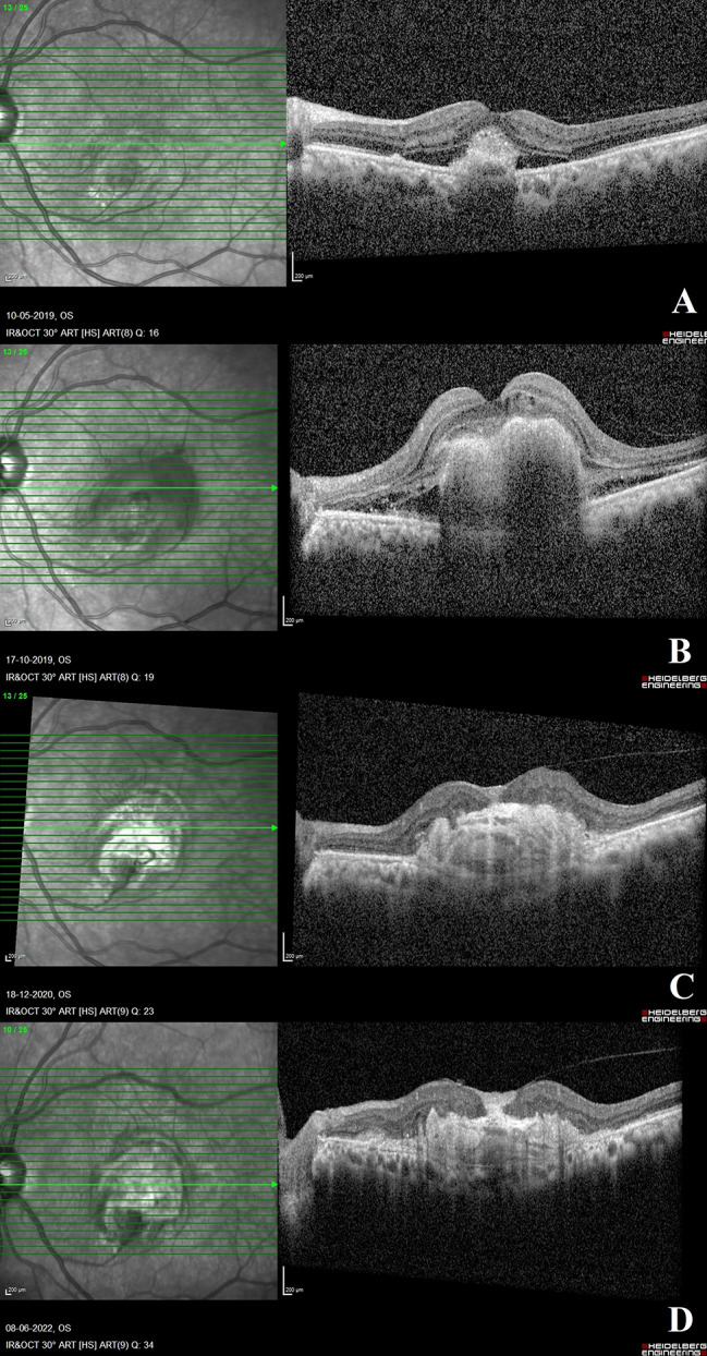
Results: We identified 530 eyes in 273 patients with chronic predominantly scarred macular neovascularisation (MNV), and 32 eyes in 29 patients revealed CPHF, representing a prevalence of 6%. Patients had a mean age of 72.76 years. Before and during the development of CPHF, Type 2 MNV was observed in all eyes. At the time of MNV diagnosis, mean logMAR visual acuity was 1.308 ± 0.559 (20/407). The average time to develop CPHF was 27.3 months since the diagnosis of MNV. At the time of CPHF identification, the mean logMAR visual acuity was 1.498 ± 0.374 (20/630). RPE tear was observed in 6% (n = 2) of CPHF eyes. The average number of intravitreal anti-VEGF injections administered prior to the diagnosis of CPHF was 2.4 and after the onset of CPHF was 0.9. The final visual acuity was not significantly different at the final follow-up visit [1.304 ± 0.42 (20/402); p = 0.646].
Conclusion: Rarely observed in eyes with predominantly scarred subfoveal type 2 MNVs and extensive subretinal fibrosis, CPHF is associated with poor visual outcomes. Its presence could possibly suggest a profibrotic effect of MNV on the posterior hyaloid.
期刊介绍:
International Journal of Retina and Vitreous focuses on the ophthalmic subspecialty of vitreoretinal disorders. The journal presents original articles on new approaches to diagnosis, outcomes of clinical trials, innovations in pharmacological therapy and surgical techniques, as well as basic science advances that impact clinical practice. Topical areas include, but are not limited to: -Imaging of the retina, choroid and vitreous -Innovations in optical coherence tomography (OCT) -Small-gauge vitrectomy, retinal detachment, chromovitrectomy -Electroretinography (ERG), microperimetry, other functional tests -Intraocular tumors -Retinal pharmacotherapy & drug delivery -Diabetic retinopathy & other vascular diseases -Age-related macular degeneration (AMD) & other macular entities

 求助内容:
求助内容: 应助结果提醒方式:


