Sang Woo Han, Jiye Kim, Sug Won Kim, Minseob Eom, Chae Eun Yang
{"title":"咀嚼间隙肌内表皮囊肿1例。","authors":"Sang Woo Han, Jiye Kim, Sug Won Kim, Minseob Eom, Chae Eun Yang","doi":"10.7181/acfs.2023.00136","DOIUrl":null,"url":null,"abstract":"<p><p>An epidermal cyst, also known as an epidermoid cyst or epidermal inclusion cyst, is the most prevalent type of cutaneous cyst. This noncancerous lesion can appear anywhere on the body, typically presenting as an asymptomatic dermal nodule with a visible central punctum. In the case presented herein, an epidermal cyst with uncommon features was misdiagnosed as a lymphatic malformation based on preoperative magnetic resonance imaging (MRI). A 61-year-old man came to us with a swollen left cheek that had been present for 11 months. The preoperative MRI revealed a 3 × 3.8 × 4.6 cm lobulated cystic lesion with thin rim enhancement in the left masticator space. The initial differential diagnosis pointed toward a lymphatic malformation. We proceeded with surgical excision of the lesion via an intraoral approach, and the specimen was sent to the pathology department. The pathological diagnosis revealed a ruptured epidermal cyst, indicating that the initial diagnosis of a lymphatic malformation based on preoperative MRI was incorrect. Epidermal cysts located under the muscle with no visible central punctum are uncommon, but should be considered if a patient presents with facial swelling.</p>","PeriodicalId":52238,"journal":{"name":"Archives of Craniofacial Surgery","volume":"24 4","pages":"193-197"},"PeriodicalIF":0.0000,"publicationDate":"2023-08-01","publicationTypes":"Journal Article","fieldsOfStudy":null,"isOpenAccess":false,"openAccessPdf":"https://ftp.ncbi.nlm.nih.gov/pub/pmc/oa_pdf/7e/ee/acfs-2023-00136.PMC10475696.pdf","citationCount":"0","resultStr":"{\"title\":\"Intramuscular epidermal cyst in the masticator space: a case report.\",\"authors\":\"Sang Woo Han, Jiye Kim, Sug Won Kim, Minseob Eom, Chae Eun Yang\",\"doi\":\"10.7181/acfs.2023.00136\",\"DOIUrl\":null,\"url\":null,\"abstract\":\"<p><p>An epidermal cyst, also known as an epidermoid cyst or epidermal inclusion cyst, is the most prevalent type of cutaneous cyst. This noncancerous lesion can appear anywhere on the body, typically presenting as an asymptomatic dermal nodule with a visible central punctum. In the case presented herein, an epidermal cyst with uncommon features was misdiagnosed as a lymphatic malformation based on preoperative magnetic resonance imaging (MRI). A 61-year-old man came to us with a swollen left cheek that had been present for 11 months. The preoperative MRI revealed a 3 × 3.8 × 4.6 cm lobulated cystic lesion with thin rim enhancement in the left masticator space. The initial differential diagnosis pointed toward a lymphatic malformation. We proceeded with surgical excision of the lesion via an intraoral approach, and the specimen was sent to the pathology department. The pathological diagnosis revealed a ruptured epidermal cyst, indicating that the initial diagnosis of a lymphatic malformation based on preoperative MRI was incorrect. Epidermal cysts located under the muscle with no visible central punctum are uncommon, but should be considered if a patient presents with facial swelling.</p>\",\"PeriodicalId\":52238,\"journal\":{\"name\":\"Archives of Craniofacial Surgery\",\"volume\":\"24 4\",\"pages\":\"193-197\"},\"PeriodicalIF\":0.0000,\"publicationDate\":\"2023-08-01\",\"publicationTypes\":\"Journal Article\",\"fieldsOfStudy\":null,\"isOpenAccess\":false,\"openAccessPdf\":\"https://ftp.ncbi.nlm.nih.gov/pub/pmc/oa_pdf/7e/ee/acfs-2023-00136.PMC10475696.pdf\",\"citationCount\":\"0\",\"resultStr\":null,\"platform\":\"Semanticscholar\",\"paperid\":null,\"PeriodicalName\":\"Archives of Craniofacial Surgery\",\"FirstCategoryId\":\"1085\",\"ListUrlMain\":\"https://doi.org/10.7181/acfs.2023.00136\",\"RegionNum\":0,\"RegionCategory\":null,\"ArticlePicture\":[],\"TitleCN\":null,\"AbstractTextCN\":null,\"PMCID\":null,\"EPubDate\":\"\",\"PubModel\":\"\",\"JCR\":\"Q2\",\"JCRName\":\"Medicine\",\"Score\":null,\"Total\":0}","platform":"Semanticscholar","paperid":null,"PeriodicalName":"Archives of Craniofacial Surgery","FirstCategoryId":"1085","ListUrlMain":"https://doi.org/10.7181/acfs.2023.00136","RegionNum":0,"RegionCategory":null,"ArticlePicture":[],"TitleCN":null,"AbstractTextCN":null,"PMCID":null,"EPubDate":"","PubModel":"","JCR":"Q2","JCRName":"Medicine","Score":null,"Total":0}
Intramuscular epidermal cyst in the masticator space: a case report.
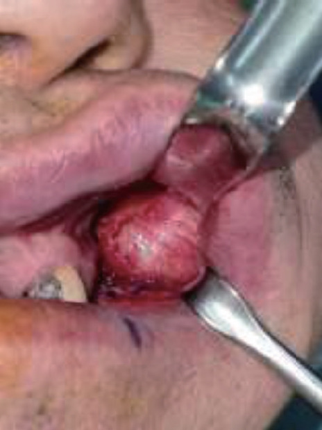
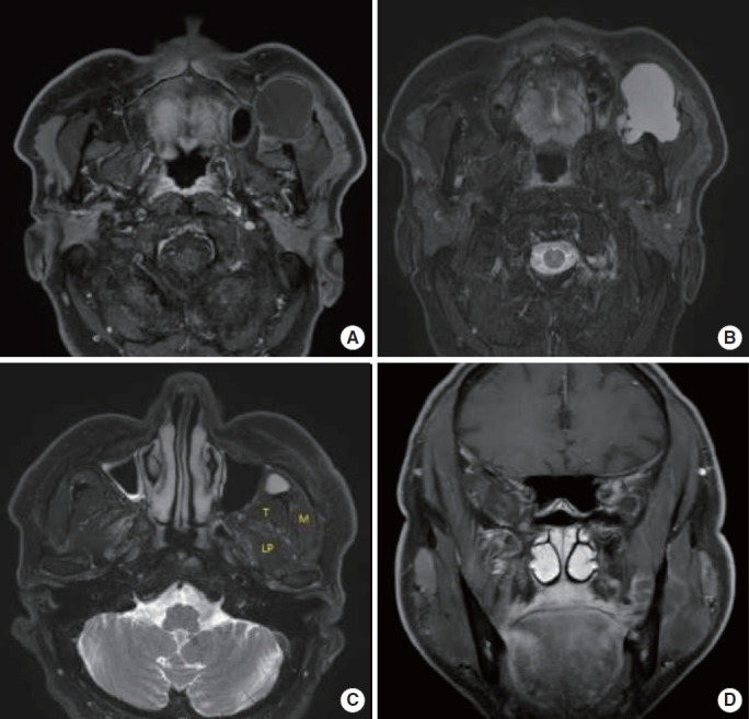
An epidermal cyst, also known as an epidermoid cyst or epidermal inclusion cyst, is the most prevalent type of cutaneous cyst. This noncancerous lesion can appear anywhere on the body, typically presenting as an asymptomatic dermal nodule with a visible central punctum. In the case presented herein, an epidermal cyst with uncommon features was misdiagnosed as a lymphatic malformation based on preoperative magnetic resonance imaging (MRI). A 61-year-old man came to us with a swollen left cheek that had been present for 11 months. The preoperative MRI revealed a 3 × 3.8 × 4.6 cm lobulated cystic lesion with thin rim enhancement in the left masticator space. The initial differential diagnosis pointed toward a lymphatic malformation. We proceeded with surgical excision of the lesion via an intraoral approach, and the specimen was sent to the pathology department. The pathological diagnosis revealed a ruptured epidermal cyst, indicating that the initial diagnosis of a lymphatic malformation based on preoperative MRI was incorrect. Epidermal cysts located under the muscle with no visible central punctum are uncommon, but should be considered if a patient presents with facial swelling.


 求助内容:
求助内容: 应助结果提醒方式:


