Amanda Leal Ferreira, Nasle Domingues Dibe, Bruna Rodrigues de Paiva, Elyzabeth Avvad Portari, Dione Corrêa de Araújo Dock, Nilma Valéria Caldeira Ferreira, Saint Clair Gomes Junior, Fábio Bastos Russomano, Cecília Vianna de Andrade
{"title":"宫颈上皮内肿瘤2级活检:p16INK4a和Ki-67生物标志物是否有助于决定治疗?横断面研究。","authors":"Amanda Leal Ferreira, Nasle Domingues Dibe, Bruna Rodrigues de Paiva, Elyzabeth Avvad Portari, Dione Corrêa de Araújo Dock, Nilma Valéria Caldeira Ferreira, Saint Clair Gomes Junior, Fábio Bastos Russomano, Cecília Vianna de Andrade","doi":"10.1590/1516-3180.2022.0527.R2.280423","DOIUrl":null,"url":null,"abstract":"<p><strong>Background: </strong>Managing cervical intraepithelial neoplasia grade 2 (CIN2) is challenging, considering the CIN2 regression rate, perinatal risks associated with excisional procedures, and insufficient well-established risk factors to predict progression.</p><p><strong>Objectives: </strong>To determine the ability of p16INK4a and Ki-67 staining in biopsies diagnosed with CIN2 to identify patients with higher-grade lesions (CIN3 or carcinoma).</p><p><strong>Design and setting: </strong>Cross-sectional study conducted at a referral center for treating uterine cervical lesions.</p><p><strong>Methods: </strong>In 79 women, we analyzed the correlation of p16INK4a and Ki-67 expression in CIN2 biopsies with the presence of a higher-grade lesions, as determined via histopathology in surgical specimens from treated women or via two colposcopies and two cytological tests during follow-up for untreated women with at least a 6-month interval. The expression of these two biomarkers was verified by at least two independent pathologists and quantified using digital algorithms.</p><p><strong>Results: </strong>Thirteen (16.8%) women with CIN2 biopsy exhibited higher-grade lesions on the surgical excision specimen or during follow-up. p16INK4a expression positively and negatively predicted the presence of higher-grade lesions in 17.19% and 86.67% patients, respectively. Ki-67 expression positively and negatively predicted the presence of higher-grade lesions in 40% and 88.24% patients, respectively.</p><p><strong>Conclusions: </strong>Negative p16INK4a and Ki67 immunohistochemical staining can assure absence of a higher-grade lesion in more than 85% of patients with CIN2 biopsies and can be used to prevent overtreatment of these patients. Positive IHC staining for p16INK4a and Ki-67 did not predict CIN3 in patients with CIN2 biopsies.</p>","PeriodicalId":49574,"journal":{"name":"Sao Paulo Medical Journal","volume":"142 1","pages":"e2022527"},"PeriodicalIF":1.3000,"publicationDate":"2023-08-25","publicationTypes":"Journal Article","fieldsOfStudy":null,"isOpenAccess":false,"openAccessPdf":"https://www.ncbi.nlm.nih.gov/pmc/articles/PMC10452003/pdf/","citationCount":"0","resultStr":"{\"title\":\"Cervical Intraepithelial Neoplasia grade 2 biopsy: Do p16INK4a and Ki-67 biomarkers contribute to the decision to treat? A cross-sectional study.\",\"authors\":\"Amanda Leal Ferreira, Nasle Domingues Dibe, Bruna Rodrigues de Paiva, Elyzabeth Avvad Portari, Dione Corrêa de Araújo Dock, Nilma Valéria Caldeira Ferreira, Saint Clair Gomes Junior, Fábio Bastos Russomano, Cecília Vianna de Andrade\",\"doi\":\"10.1590/1516-3180.2022.0527.R2.280423\",\"DOIUrl\":null,\"url\":null,\"abstract\":\"<p><strong>Background: </strong>Managing cervical intraepithelial neoplasia grade 2 (CIN2) is challenging, considering the CIN2 regression rate, perinatal risks associated with excisional procedures, and insufficient well-established risk factors to predict progression.</p><p><strong>Objectives: </strong>To determine the ability of p16INK4a and Ki-67 staining in biopsies diagnosed with CIN2 to identify patients with higher-grade lesions (CIN3 or carcinoma).</p><p><strong>Design and setting: </strong>Cross-sectional study conducted at a referral center for treating uterine cervical lesions.</p><p><strong>Methods: </strong>In 79 women, we analyzed the correlation of p16INK4a and Ki-67 expression in CIN2 biopsies with the presence of a higher-grade lesions, as determined via histopathology in surgical specimens from treated women or via two colposcopies and two cytological tests during follow-up for untreated women with at least a 6-month interval. The expression of these two biomarkers was verified by at least two independent pathologists and quantified using digital algorithms.</p><p><strong>Results: </strong>Thirteen (16.8%) women with CIN2 biopsy exhibited higher-grade lesions on the surgical excision specimen or during follow-up. p16INK4a expression positively and negatively predicted the presence of higher-grade lesions in 17.19% and 86.67% patients, respectively. Ki-67 expression positively and negatively predicted the presence of higher-grade lesions in 40% and 88.24% patients, respectively.</p><p><strong>Conclusions: </strong>Negative p16INK4a and Ki67 immunohistochemical staining can assure absence of a higher-grade lesion in more than 85% of patients with CIN2 biopsies and can be used to prevent overtreatment of these patients. Positive IHC staining for p16INK4a and Ki-67 did not predict CIN3 in patients with CIN2 biopsies.</p>\",\"PeriodicalId\":49574,\"journal\":{\"name\":\"Sao Paulo Medical Journal\",\"volume\":\"142 1\",\"pages\":\"e2022527\"},\"PeriodicalIF\":1.3000,\"publicationDate\":\"2023-08-25\",\"publicationTypes\":\"Journal Article\",\"fieldsOfStudy\":null,\"isOpenAccess\":false,\"openAccessPdf\":\"https://www.ncbi.nlm.nih.gov/pmc/articles/PMC10452003/pdf/\",\"citationCount\":\"0\",\"resultStr\":null,\"platform\":\"Semanticscholar\",\"paperid\":null,\"PeriodicalName\":\"Sao Paulo Medical Journal\",\"FirstCategoryId\":\"3\",\"ListUrlMain\":\"https://doi.org/10.1590/1516-3180.2022.0527.R2.280423\",\"RegionNum\":4,\"RegionCategory\":\"医学\",\"ArticlePicture\":[],\"TitleCN\":null,\"AbstractTextCN\":null,\"PMCID\":null,\"EPubDate\":\"2023/1/1 0:00:00\",\"PubModel\":\"eCollection\",\"JCR\":\"Q2\",\"JCRName\":\"MEDICINE, GENERAL & INTERNAL\",\"Score\":null,\"Total\":0}","platform":"Semanticscholar","paperid":null,"PeriodicalName":"Sao Paulo Medical Journal","FirstCategoryId":"3","ListUrlMain":"https://doi.org/10.1590/1516-3180.2022.0527.R2.280423","RegionNum":4,"RegionCategory":"医学","ArticlePicture":[],"TitleCN":null,"AbstractTextCN":null,"PMCID":null,"EPubDate":"2023/1/1 0:00:00","PubModel":"eCollection","JCR":"Q2","JCRName":"MEDICINE, GENERAL & INTERNAL","Score":null,"Total":0}
Cervical Intraepithelial Neoplasia grade 2 biopsy: Do p16INK4a and Ki-67 biomarkers contribute to the decision to treat? A cross-sectional study.
Background: Managing cervical intraepithelial neoplasia grade 2 (CIN2) is challenging, considering the CIN2 regression rate, perinatal risks associated with excisional procedures, and insufficient well-established risk factors to predict progression.
Objectives: To determine the ability of p16INK4a and Ki-67 staining in biopsies diagnosed with CIN2 to identify patients with higher-grade lesions (CIN3 or carcinoma).
Design and setting: Cross-sectional study conducted at a referral center for treating uterine cervical lesions.
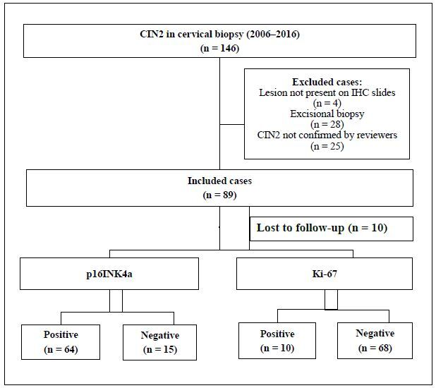
Methods: In 79 women, we analyzed the correlation of p16INK4a and Ki-67 expression in CIN2 biopsies with the presence of a higher-grade lesions, as determined via histopathology in surgical specimens from treated women or via two colposcopies and two cytological tests during follow-up for untreated women with at least a 6-month interval. The expression of these two biomarkers was verified by at least two independent pathologists and quantified using digital algorithms.
Results: Thirteen (16.8%) women with CIN2 biopsy exhibited higher-grade lesions on the surgical excision specimen or during follow-up. p16INK4a expression positively and negatively predicted the presence of higher-grade lesions in 17.19% and 86.67% patients, respectively. Ki-67 expression positively and negatively predicted the presence of higher-grade lesions in 40% and 88.24% patients, respectively.
Conclusions: Negative p16INK4a and Ki67 immunohistochemical staining can assure absence of a higher-grade lesion in more than 85% of patients with CIN2 biopsies and can be used to prevent overtreatment of these patients. Positive IHC staining for p16INK4a and Ki-67 did not predict CIN3 in patients with CIN2 biopsies.
期刊介绍:
Published bimonthly by the Associação Paulista de Medicina, the journal accepts articles in the fields of clinical health science (internal medicine, gynecology and obstetrics, mental health, surgery, pediatrics and public health). Articles will be accepted in the form of original articles (clinical trials, cohort, case-control, prevalence, incidence, accuracy and cost-effectiveness studies and systematic reviews with or without meta-analysis), narrative reviews of the literature, case reports, short communications and letters to the editor. Papers with a commercial objective will not be accepted.
 求助内容:
求助内容: 应助结果提醒方式:


