Jason Langley, Kristy S Hwang, Daniel E Huddleston, Xiaoping P Hu
{"title":"前驱、早期和中度帕金森病患者的黑质体积损失。","authors":"Jason Langley, Kristy S Hwang, Daniel E Huddleston, Xiaoping P Hu","doi":"10.1101/2023.08.19.23294281","DOIUrl":null,"url":null,"abstract":"<p><p>The loss of melanized neurons in the substantia nigra pars compacta (SNc) is a hallmark pathology in Parkinson's disease (PD). Melanized neurons in SNc can be visualized in vivo using magnetization transfer (MT) effects. Nigral volume was extracted in data acquired with a MT-prepared gradient echo sequence in 50 controls, 90 non-manifest carriers (46 LRRK2 and 44 GBA1 nonmanifest carriers), 217 prodromal hyposmic participants, 76 participants with rapid eye movement sleep behavior disorder (RBD), 194 de novo PD patients and 26 moderate PD patients from the Parkinson's Progressive Markers Initiative. No difference in nigral volume was seen between controls and LRRK2 and GBA1 non-manifest carriers ( <i>F</i> =0.732; <i>P</i> =0.483). A significant main effect in group was observed between controls, prodromal hyposmic participants, RBD participants, and overt PD patients ( <i>F</i> =9.882; <i>P</i> <10 <sup>-3</sup> ). This study shows that nigral depigmentation can be robustly detected in prodromal and overt PD populations.</p>","PeriodicalId":18659,"journal":{"name":"medRxiv : the preprint server for health sciences","volume":" ","pages":""},"PeriodicalIF":0.0000,"publicationDate":"2025-03-21","publicationTypes":"Journal Article","fieldsOfStudy":null,"isOpenAccess":false,"openAccessPdf":"https://www.ncbi.nlm.nih.gov/pmc/articles/PMC10462207/pdf/","citationCount":"0","resultStr":"{\"title\":\"Nigral volume loss in prodromal, early, and moderate Parkinson's disease.\",\"authors\":\"Jason Langley, Kristy S Hwang, Daniel E Huddleston, Xiaoping P Hu\",\"doi\":\"10.1101/2023.08.19.23294281\",\"DOIUrl\":null,\"url\":null,\"abstract\":\"<p><p>The loss of melanized neurons in the substantia nigra pars compacta (SNc) is a hallmark pathology in Parkinson's disease (PD). Melanized neurons in SNc can be visualized in vivo using magnetization transfer (MT) effects. Nigral volume was extracted in data acquired with a MT-prepared gradient echo sequence in 50 controls, 90 non-manifest carriers (46 LRRK2 and 44 GBA1 nonmanifest carriers), 217 prodromal hyposmic participants, 76 participants with rapid eye movement sleep behavior disorder (RBD), 194 de novo PD patients and 26 moderate PD patients from the Parkinson's Progressive Markers Initiative. No difference in nigral volume was seen between controls and LRRK2 and GBA1 non-manifest carriers ( <i>F</i> =0.732; <i>P</i> =0.483). A significant main effect in group was observed between controls, prodromal hyposmic participants, RBD participants, and overt PD patients ( <i>F</i> =9.882; <i>P</i> <10 <sup>-3</sup> ). This study shows that nigral depigmentation can be robustly detected in prodromal and overt PD populations.</p>\",\"PeriodicalId\":18659,\"journal\":{\"name\":\"medRxiv : the preprint server for health sciences\",\"volume\":\" \",\"pages\":\"\"},\"PeriodicalIF\":0.0000,\"publicationDate\":\"2025-03-21\",\"publicationTypes\":\"Journal Article\",\"fieldsOfStudy\":null,\"isOpenAccess\":false,\"openAccessPdf\":\"https://www.ncbi.nlm.nih.gov/pmc/articles/PMC10462207/pdf/\",\"citationCount\":\"0\",\"resultStr\":null,\"platform\":\"Semanticscholar\",\"paperid\":null,\"PeriodicalName\":\"medRxiv : the preprint server for health sciences\",\"FirstCategoryId\":\"1085\",\"ListUrlMain\":\"https://doi.org/10.1101/2023.08.19.23294281\",\"RegionNum\":0,\"RegionCategory\":null,\"ArticlePicture\":[],\"TitleCN\":null,\"AbstractTextCN\":null,\"PMCID\":null,\"EPubDate\":\"\",\"PubModel\":\"\",\"JCR\":\"\",\"JCRName\":\"\",\"Score\":null,\"Total\":0}","platform":"Semanticscholar","paperid":null,"PeriodicalName":"medRxiv : the preprint server for health sciences","FirstCategoryId":"1085","ListUrlMain":"https://doi.org/10.1101/2023.08.19.23294281","RegionNum":0,"RegionCategory":null,"ArticlePicture":[],"TitleCN":null,"AbstractTextCN":null,"PMCID":null,"EPubDate":"","PubModel":"","JCR":"","JCRName":"","Score":null,"Total":0}
Nigral volume loss in prodromal, early, and moderate Parkinson's disease.
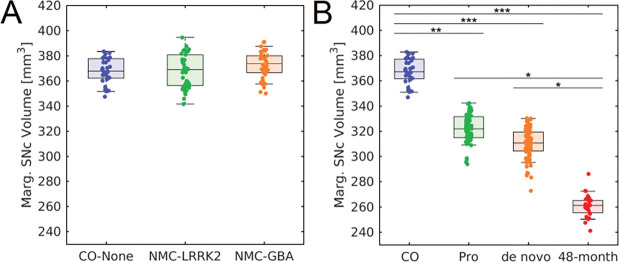
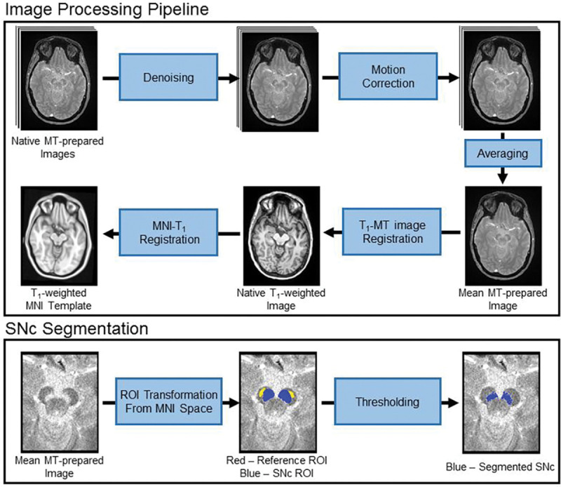
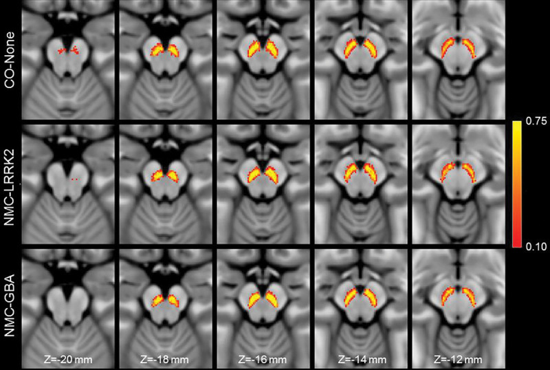
The loss of melanized neurons in the substantia nigra pars compacta (SNc) is a hallmark pathology in Parkinson's disease (PD). Melanized neurons in SNc can be visualized in vivo using magnetization transfer (MT) effects. Nigral volume was extracted in data acquired with a MT-prepared gradient echo sequence in 50 controls, 90 non-manifest carriers (46 LRRK2 and 44 GBA1 nonmanifest carriers), 217 prodromal hyposmic participants, 76 participants with rapid eye movement sleep behavior disorder (RBD), 194 de novo PD patients and 26 moderate PD patients from the Parkinson's Progressive Markers Initiative. No difference in nigral volume was seen between controls and LRRK2 and GBA1 non-manifest carriers ( F =0.732; P =0.483). A significant main effect in group was observed between controls, prodromal hyposmic participants, RBD participants, and overt PD patients ( F =9.882; P <10 -3 ). This study shows that nigral depigmentation can be robustly detected in prodromal and overt PD populations.

 求助内容:
求助内容: 应助结果提醒方式:


