Fatemeh Abbasi Yeganeh, Corinne Summerill, Zhongjun Hu, Hamidreza Rahmani, Dianne W Taylor, Kenneth A Taylor
{"title":"分离的印度小尾丝虫Z盘的冷冻电镜三维图像重建。","authors":"Fatemeh Abbasi Yeganeh, Corinne Summerill, Zhongjun Hu, Hamidreza Rahmani, Dianne W Taylor, Kenneth A Taylor","doi":"10.1007/s10974-023-09657-1","DOIUrl":null,"url":null,"abstract":"<p><p>The Z-disk of striated muscle defines the ends of the sarcomere, which repeats many times within the muscle fiber. Here we report application of cryoelectron tomography and subtomogram averaging to Z-disks isolated from the flight muscles of the large waterbug Lethocerus indicus. We use high salt solutions to remove the myosin containing filaments and use gelsolin to remove the actin filaments of the A- and I-bands leaving only the thin filaments within the Z-disk which were then frozen for cryoelectron microscopy. The Lethocerus Z-disk structure is similar in many ways to the previously studied Z-disk of the honeybee Apis mellifera. At the corners of the unit cell are positioned trimers of paired antiparallel F-actins defining a large solvent channel, whereas at the trigonal positions are positioned F-actin trimers converging slowly towards their (+) ends defining a small solvent channel through the Z-disk. These near parallel F-actins terminate at different Z-heights within the Z-disk. The two types of solvent channel in Lethocerus are similar in size compared to those of Apis which are very different in size. Two types of α-actinin crosslinks were observed between oppositely oriented actin filaments. In one of these, the α-actinin long axis is almost parallel to the F-actins it crosslinks. In the other, the α-actinins are at a small but distinctive angle with respect to the crosslinked actin filaments. The utility of isolated Z-disks for structure determination is discussed.</p>","PeriodicalId":16422,"journal":{"name":"Journal of Muscle Research and Cell Motility","volume":" ","pages":"271-286"},"PeriodicalIF":1.7000,"publicationDate":"2023-12-01","publicationTypes":"Journal Article","fieldsOfStudy":null,"isOpenAccess":false,"openAccessPdf":"https://www.ncbi.nlm.nih.gov/pmc/articles/PMC10843718/pdf/","citationCount":"0","resultStr":"{\"title\":\"The cryo-EM 3D image reconstruction of isolated Lethocerus indicus Z-discs.\",\"authors\":\"Fatemeh Abbasi Yeganeh, Corinne Summerill, Zhongjun Hu, Hamidreza Rahmani, Dianne W Taylor, Kenneth A Taylor\",\"doi\":\"10.1007/s10974-023-09657-1\",\"DOIUrl\":null,\"url\":null,\"abstract\":\"<p><p>The Z-disk of striated muscle defines the ends of the sarcomere, which repeats many times within the muscle fiber. Here we report application of cryoelectron tomography and subtomogram averaging to Z-disks isolated from the flight muscles of the large waterbug Lethocerus indicus. We use high salt solutions to remove the myosin containing filaments and use gelsolin to remove the actin filaments of the A- and I-bands leaving only the thin filaments within the Z-disk which were then frozen for cryoelectron microscopy. The Lethocerus Z-disk structure is similar in many ways to the previously studied Z-disk of the honeybee Apis mellifera. At the corners of the unit cell are positioned trimers of paired antiparallel F-actins defining a large solvent channel, whereas at the trigonal positions are positioned F-actin trimers converging slowly towards their (+) ends defining a small solvent channel through the Z-disk. These near parallel F-actins terminate at different Z-heights within the Z-disk. The two types of solvent channel in Lethocerus are similar in size compared to those of Apis which are very different in size. Two types of α-actinin crosslinks were observed between oppositely oriented actin filaments. In one of these, the α-actinin long axis is almost parallel to the F-actins it crosslinks. In the other, the α-actinins are at a small but distinctive angle with respect to the crosslinked actin filaments. The utility of isolated Z-disks for structure determination is discussed.</p>\",\"PeriodicalId\":16422,\"journal\":{\"name\":\"Journal of Muscle Research and Cell Motility\",\"volume\":\" \",\"pages\":\"271-286\"},\"PeriodicalIF\":1.7000,\"publicationDate\":\"2023-12-01\",\"publicationTypes\":\"Journal Article\",\"fieldsOfStudy\":null,\"isOpenAccess\":false,\"openAccessPdf\":\"https://www.ncbi.nlm.nih.gov/pmc/articles/PMC10843718/pdf/\",\"citationCount\":\"0\",\"resultStr\":null,\"platform\":\"Semanticscholar\",\"paperid\":null,\"PeriodicalName\":\"Journal of Muscle Research and Cell Motility\",\"FirstCategoryId\":\"99\",\"ListUrlMain\":\"https://doi.org/10.1007/s10974-023-09657-1\",\"RegionNum\":3,\"RegionCategory\":\"生物学\",\"ArticlePicture\":[],\"TitleCN\":null,\"AbstractTextCN\":null,\"PMCID\":null,\"EPubDate\":\"2023/9/3 0:00:00\",\"PubModel\":\"Epub\",\"JCR\":\"Q4\",\"JCRName\":\"CELL BIOLOGY\",\"Score\":null,\"Total\":0}","platform":"Semanticscholar","paperid":null,"PeriodicalName":"Journal of Muscle Research and Cell Motility","FirstCategoryId":"99","ListUrlMain":"https://doi.org/10.1007/s10974-023-09657-1","RegionNum":3,"RegionCategory":"生物学","ArticlePicture":[],"TitleCN":null,"AbstractTextCN":null,"PMCID":null,"EPubDate":"2023/9/3 0:00:00","PubModel":"Epub","JCR":"Q4","JCRName":"CELL BIOLOGY","Score":null,"Total":0}
The cryo-EM 3D image reconstruction of isolated Lethocerus indicus Z-discs.
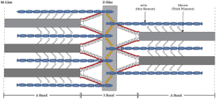
The Z-disk of striated muscle defines the ends of the sarcomere, which repeats many times within the muscle fiber. Here we report application of cryoelectron tomography and subtomogram averaging to Z-disks isolated from the flight muscles of the large waterbug Lethocerus indicus. We use high salt solutions to remove the myosin containing filaments and use gelsolin to remove the actin filaments of the A- and I-bands leaving only the thin filaments within the Z-disk which were then frozen for cryoelectron microscopy. The Lethocerus Z-disk structure is similar in many ways to the previously studied Z-disk of the honeybee Apis mellifera. At the corners of the unit cell are positioned trimers of paired antiparallel F-actins defining a large solvent channel, whereas at the trigonal positions are positioned F-actin trimers converging slowly towards their (+) ends defining a small solvent channel through the Z-disk. These near parallel F-actins terminate at different Z-heights within the Z-disk. The two types of solvent channel in Lethocerus are similar in size compared to those of Apis which are very different in size. Two types of α-actinin crosslinks were observed between oppositely oriented actin filaments. In one of these, the α-actinin long axis is almost parallel to the F-actins it crosslinks. In the other, the α-actinins are at a small but distinctive angle with respect to the crosslinked actin filaments. The utility of isolated Z-disks for structure determination is discussed.
期刊介绍:
The Journal of Muscle Research and Cell Motility has as its main aim the publication of original research which bears on either the excitation and contraction of muscle, the analysis of any one of the processes involved therein, the processes underlying contractility and motility of animal and plant cells, the toxicology and pharmacology related to contractility, or the formation, dynamics and turnover of contractile structures in muscle and non-muscle cells. Studies describing the impact of pathogenic mutations in genes encoding components of contractile structures in humans or animals are welcome, provided they offer mechanistic insight into the disease process or the underlying gene function. The policy of the Journal is to encourage any form of novel practical study whatever its specialist interest, as long as it falls within this broad field. Theoretical essays are welcome provided that they are concise and suggest practical ways in which they may be tested. Manuscripts reporting new mutations in known disease genes without validation and mechanistic insight will not be considered. It is the policy of the journal that cells lines, hybridomas and DNA clones should be made available by the developers to any qualified investigator. Submission of a manuscript for publication constitutes an agreement of the authors to abide by this principle.
 求助内容:
求助内容: 应助结果提醒方式:


