Doo Sik Park, Eun Hyun Cho, Kyung Hoon Park, Soo Min Jo, Bumjung Park, Sun Huh
{"title":"在韩国生活的中国妇女声带舌口病的病理切片诊断。","authors":"Doo Sik Park, Eun Hyun Cho, Kyung Hoon Park, Soo Min Jo, Bumjung Park, Sun Huh","doi":"10.3347/PHD.23065","DOIUrl":null,"url":null,"abstract":"<p><p>This study aimed to describe a rare case of gnathostomiasis in the vocal cord. A 54-year-old Chinese woman living in Korea visited with a chief complaint of voice change at the outpatient department of otorhinolaryngology in Hallym Sacred Heart Hospital, Hallym University on August 2, 2021. She had eaten raw conger a few weeks before the voice change developed, but her medical history and physical examinations demonstrated neither gastrointestinal symptoms nor other health problems. A round and red cystic lesion, recognized in the anterior part of the right vocal cord, was removed using forceps and scissors through laryngeal microsurgery. The histopathological specimen of the cyst revealed 3 cross-sections of a nematode larva in the lumen of the cyst wall composed of inflammatory cells and fibrotic tissues. They differ in diameter, from 190 μm to 235 μm. They showed characteristic cuticular layers with tegumental spines, somatic muscle layers, and gastrointestinal tracts such as the esophagus and intestine. Notably, intestinal sections consisted of 27-28 lining cells containing 0-4 nuclei per cell. We tentatively identified the nematode larva recovered from the vocal cord cystic lesion as the third-stage larva of Gnathostoma, probably G. nipponicum or G. hispidum, based on the sectional morphologies.</p>","PeriodicalId":74397,"journal":{"name":"Parasites, hosts and diseases","volume":"61 3","pages":"298-303"},"PeriodicalIF":1.3000,"publicationDate":"2023-08-01","publicationTypes":"Journal Article","fieldsOfStudy":null,"isOpenAccess":false,"openAccessPdf":"https://ftp.ncbi.nlm.nih.gov/pub/pmc/oa_pdf/4e/a0/phd-23065.PMC10471466.pdf","citationCount":"0","resultStr":"{\"title\":\"A case of vocal cord gnathostomiasis diagnosed with sectional morphologies in a histopathological specimen from a Chinese woman living in Korea.\",\"authors\":\"Doo Sik Park, Eun Hyun Cho, Kyung Hoon Park, Soo Min Jo, Bumjung Park, Sun Huh\",\"doi\":\"10.3347/PHD.23065\",\"DOIUrl\":null,\"url\":null,\"abstract\":\"<p><p>This study aimed to describe a rare case of gnathostomiasis in the vocal cord. A 54-year-old Chinese woman living in Korea visited with a chief complaint of voice change at the outpatient department of otorhinolaryngology in Hallym Sacred Heart Hospital, Hallym University on August 2, 2021. She had eaten raw conger a few weeks before the voice change developed, but her medical history and physical examinations demonstrated neither gastrointestinal symptoms nor other health problems. A round and red cystic lesion, recognized in the anterior part of the right vocal cord, was removed using forceps and scissors through laryngeal microsurgery. The histopathological specimen of the cyst revealed 3 cross-sections of a nematode larva in the lumen of the cyst wall composed of inflammatory cells and fibrotic tissues. They differ in diameter, from 190 μm to 235 μm. They showed characteristic cuticular layers with tegumental spines, somatic muscle layers, and gastrointestinal tracts such as the esophagus and intestine. Notably, intestinal sections consisted of 27-28 lining cells containing 0-4 nuclei per cell. We tentatively identified the nematode larva recovered from the vocal cord cystic lesion as the third-stage larva of Gnathostoma, probably G. nipponicum or G. hispidum, based on the sectional morphologies.</p>\",\"PeriodicalId\":74397,\"journal\":{\"name\":\"Parasites, hosts and diseases\",\"volume\":\"61 3\",\"pages\":\"298-303\"},\"PeriodicalIF\":1.3000,\"publicationDate\":\"2023-08-01\",\"publicationTypes\":\"Journal Article\",\"fieldsOfStudy\":null,\"isOpenAccess\":false,\"openAccessPdf\":\"https://ftp.ncbi.nlm.nih.gov/pub/pmc/oa_pdf/4e/a0/phd-23065.PMC10471466.pdf\",\"citationCount\":\"0\",\"resultStr\":null,\"platform\":\"Semanticscholar\",\"paperid\":null,\"PeriodicalName\":\"Parasites, hosts and diseases\",\"FirstCategoryId\":\"1085\",\"ListUrlMain\":\"https://doi.org/10.3347/PHD.23065\",\"RegionNum\":0,\"RegionCategory\":null,\"ArticlePicture\":[],\"TitleCN\":null,\"AbstractTextCN\":null,\"PMCID\":null,\"EPubDate\":\"\",\"PubModel\":\"\",\"JCR\":\"0\",\"JCRName\":\"PARASITOLOGY\",\"Score\":null,\"Total\":0}","platform":"Semanticscholar","paperid":null,"PeriodicalName":"Parasites, hosts and diseases","FirstCategoryId":"1085","ListUrlMain":"https://doi.org/10.3347/PHD.23065","RegionNum":0,"RegionCategory":null,"ArticlePicture":[],"TitleCN":null,"AbstractTextCN":null,"PMCID":null,"EPubDate":"","PubModel":"","JCR":"0","JCRName":"PARASITOLOGY","Score":null,"Total":0}
A case of vocal cord gnathostomiasis diagnosed with sectional morphologies in a histopathological specimen from a Chinese woman living in Korea.
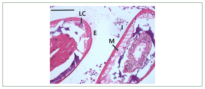
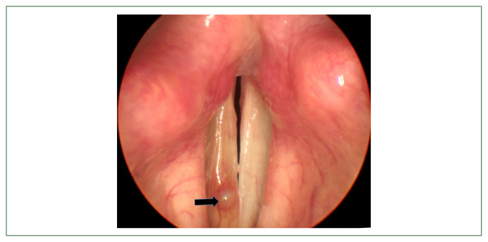
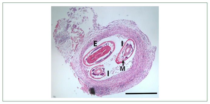
This study aimed to describe a rare case of gnathostomiasis in the vocal cord. A 54-year-old Chinese woman living in Korea visited with a chief complaint of voice change at the outpatient department of otorhinolaryngology in Hallym Sacred Heart Hospital, Hallym University on August 2, 2021. She had eaten raw conger a few weeks before the voice change developed, but her medical history and physical examinations demonstrated neither gastrointestinal symptoms nor other health problems. A round and red cystic lesion, recognized in the anterior part of the right vocal cord, was removed using forceps and scissors through laryngeal microsurgery. The histopathological specimen of the cyst revealed 3 cross-sections of a nematode larva in the lumen of the cyst wall composed of inflammatory cells and fibrotic tissues. They differ in diameter, from 190 μm to 235 μm. They showed characteristic cuticular layers with tegumental spines, somatic muscle layers, and gastrointestinal tracts such as the esophagus and intestine. Notably, intestinal sections consisted of 27-28 lining cells containing 0-4 nuclei per cell. We tentatively identified the nematode larva recovered from the vocal cord cystic lesion as the third-stage larva of Gnathostoma, probably G. nipponicum or G. hispidum, based on the sectional morphologies.

 求助内容:
求助内容: 应助结果提醒方式:


