Scott MacDonald Black, Craig Maclean, Pauline Hall Barrientos, Konstantinos Ritos, Asimina Kazakidi
{"title":"使用4D流MRI幅值图像的多个时间帧重建和验证用于计算流体动力学的动脉几何结构。","authors":"Scott MacDonald Black, Craig Maclean, Pauline Hall Barrientos, Konstantinos Ritos, Asimina Kazakidi","doi":"10.1007/s13239-023-00679-x","DOIUrl":null,"url":null,"abstract":"<p><strong>Purpose: </strong>Segmentation and reconstruction of arterial blood vessels is a fundamental step in the translation of computational fluid dynamics (CFD) to the clinical practice. Four-dimensional flow magnetic resonance imaging (4D Flow-MRI) can provide detailed information of blood flow but processing this information to elucidate the underlying anatomical structures is challenging. In this study, we present a novel approach to create high-contrast anatomical images from retrospective 4D Flow-MRI data.</p><p><strong>Methods: </strong>For healthy and clinical cases, the 3D instantaneous velocities at multiple cardiac time steps were superimposed directly onto the 4D Flow-MRI magnitude images and combined into a single composite frame. This new Composite Phase-Contrast Magnetic Resonance Angiogram (CPC-MRA) resulted in enhanced and uniform contrast within the lumen. These images were subsequently segmented and reconstructed to generate 3D arterial models for CFD. Using the time-dependent, 3D incompressible Reynolds-averaged Navier-Stokes equations, the transient aortic haemodynamics was computed within a rigid wall model of patient geometries.</p><p><strong>Results: </strong>Validation of these models against the gold standard CT-based approach showed no statistically significant inter-modality difference regarding vessel radius or curvature (p > 0.05), and a similar Dice Similarity Coefficient and Hausdorff Distance. CFD-derived near-wall hemodynamics indicated a significant inter-modality difference (p > 0.05), though these absolute errors were small. When compared to the in vivo data, CFD-derived velocities were qualitatively similar.</p><p><strong>Conclusion: </strong>This proof-of-concept study demonstrated that functional 4D Flow-MRI information can be utilized to retrospectively generate anatomical information for CFD models in the absence of standard imaging datasets and intravenous contrast.</p>","PeriodicalId":54322,"journal":{"name":"Cardiovascular Engineering and Technology","volume":" ","pages":"655-676"},"PeriodicalIF":1.6000,"publicationDate":"2023-10-01","publicationTypes":"Journal Article","fieldsOfStudy":null,"isOpenAccess":false,"openAccessPdf":"https://www.ncbi.nlm.nih.gov/pmc/articles/PMC10602980/pdf/","citationCount":"1","resultStr":"{\"title\":\"Reconstruction and Validation of Arterial Geometries for Computational Fluid Dynamics Using Multiple Temporal Frames of 4D Flow-MRI Magnitude Images.\",\"authors\":\"Scott MacDonald Black, Craig Maclean, Pauline Hall Barrientos, Konstantinos Ritos, Asimina Kazakidi\",\"doi\":\"10.1007/s13239-023-00679-x\",\"DOIUrl\":null,\"url\":null,\"abstract\":\"<p><strong>Purpose: </strong>Segmentation and reconstruction of arterial blood vessels is a fundamental step in the translation of computational fluid dynamics (CFD) to the clinical practice. Four-dimensional flow magnetic resonance imaging (4D Flow-MRI) can provide detailed information of blood flow but processing this information to elucidate the underlying anatomical structures is challenging. In this study, we present a novel approach to create high-contrast anatomical images from retrospective 4D Flow-MRI data.</p><p><strong>Methods: </strong>For healthy and clinical cases, the 3D instantaneous velocities at multiple cardiac time steps were superimposed directly onto the 4D Flow-MRI magnitude images and combined into a single composite frame. This new Composite Phase-Contrast Magnetic Resonance Angiogram (CPC-MRA) resulted in enhanced and uniform contrast within the lumen. These images were subsequently segmented and reconstructed to generate 3D arterial models for CFD. Using the time-dependent, 3D incompressible Reynolds-averaged Navier-Stokes equations, the transient aortic haemodynamics was computed within a rigid wall model of patient geometries.</p><p><strong>Results: </strong>Validation of these models against the gold standard CT-based approach showed no statistically significant inter-modality difference regarding vessel radius or curvature (p > 0.05), and a similar Dice Similarity Coefficient and Hausdorff Distance. CFD-derived near-wall hemodynamics indicated a significant inter-modality difference (p > 0.05), though these absolute errors were small. When compared to the in vivo data, CFD-derived velocities were qualitatively similar.</p><p><strong>Conclusion: </strong>This proof-of-concept study demonstrated that functional 4D Flow-MRI information can be utilized to retrospectively generate anatomical information for CFD models in the absence of standard imaging datasets and intravenous contrast.</p>\",\"PeriodicalId\":54322,\"journal\":{\"name\":\"Cardiovascular Engineering and Technology\",\"volume\":\" \",\"pages\":\"655-676\"},\"PeriodicalIF\":1.6000,\"publicationDate\":\"2023-10-01\",\"publicationTypes\":\"Journal Article\",\"fieldsOfStudy\":null,\"isOpenAccess\":false,\"openAccessPdf\":\"https://www.ncbi.nlm.nih.gov/pmc/articles/PMC10602980/pdf/\",\"citationCount\":\"1\",\"resultStr\":null,\"platform\":\"Semanticscholar\",\"paperid\":null,\"PeriodicalName\":\"Cardiovascular Engineering and Technology\",\"FirstCategoryId\":\"5\",\"ListUrlMain\":\"https://doi.org/10.1007/s13239-023-00679-x\",\"RegionNum\":4,\"RegionCategory\":\"医学\",\"ArticlePicture\":[],\"TitleCN\":null,\"AbstractTextCN\":null,\"PMCID\":null,\"EPubDate\":\"2023/8/31 0:00:00\",\"PubModel\":\"Epub\",\"JCR\":\"Q3\",\"JCRName\":\"CARDIAC & CARDIOVASCULAR SYSTEMS\",\"Score\":null,\"Total\":0}","platform":"Semanticscholar","paperid":null,"PeriodicalName":"Cardiovascular Engineering and Technology","FirstCategoryId":"5","ListUrlMain":"https://doi.org/10.1007/s13239-023-00679-x","RegionNum":4,"RegionCategory":"医学","ArticlePicture":[],"TitleCN":null,"AbstractTextCN":null,"PMCID":null,"EPubDate":"2023/8/31 0:00:00","PubModel":"Epub","JCR":"Q3","JCRName":"CARDIAC & CARDIOVASCULAR SYSTEMS","Score":null,"Total":0}
Reconstruction and Validation of Arterial Geometries for Computational Fluid Dynamics Using Multiple Temporal Frames of 4D Flow-MRI Magnitude Images.
Purpose: Segmentation and reconstruction of arterial blood vessels is a fundamental step in the translation of computational fluid dynamics (CFD) to the clinical practice. Four-dimensional flow magnetic resonance imaging (4D Flow-MRI) can provide detailed information of blood flow but processing this information to elucidate the underlying anatomical structures is challenging. In this study, we present a novel approach to create high-contrast anatomical images from retrospective 4D Flow-MRI data.
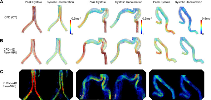
Methods: For healthy and clinical cases, the 3D instantaneous velocities at multiple cardiac time steps were superimposed directly onto the 4D Flow-MRI magnitude images and combined into a single composite frame. This new Composite Phase-Contrast Magnetic Resonance Angiogram (CPC-MRA) resulted in enhanced and uniform contrast within the lumen. These images were subsequently segmented and reconstructed to generate 3D arterial models for CFD. Using the time-dependent, 3D incompressible Reynolds-averaged Navier-Stokes equations, the transient aortic haemodynamics was computed within a rigid wall model of patient geometries.
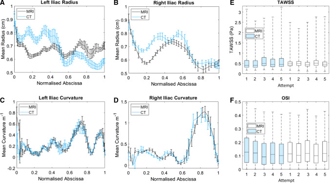
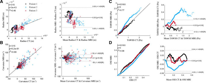
Results: Validation of these models against the gold standard CT-based approach showed no statistically significant inter-modality difference regarding vessel radius or curvature (p > 0.05), and a similar Dice Similarity Coefficient and Hausdorff Distance. CFD-derived near-wall hemodynamics indicated a significant inter-modality difference (p > 0.05), though these absolute errors were small. When compared to the in vivo data, CFD-derived velocities were qualitatively similar.
Conclusion: This proof-of-concept study demonstrated that functional 4D Flow-MRI information can be utilized to retrospectively generate anatomical information for CFD models in the absence of standard imaging datasets and intravenous contrast.
期刊介绍:
Cardiovascular Engineering and Technology is a journal publishing the spectrum of basic to translational research in all aspects of cardiovascular physiology and medical treatment. It is the forum for academic and industrial investigators to disseminate research that utilizes engineering principles and methods to advance fundamental knowledge and technological solutions related to the cardiovascular system. Manuscripts spanning from subcellular to systems level topics are invited, including but not limited to implantable medical devices, hemodynamics and tissue biomechanics, functional imaging, surgical devices, electrophysiology, tissue engineering and regenerative medicine, diagnostic instruments, transport and delivery of biologics, and sensors. In addition to manuscripts describing the original publication of research, manuscripts reviewing developments in these topics or their state-of-art are also invited.
 求助内容:
求助内容: 应助结果提醒方式:


