{"title":"植体固定物引起的鼻腔穿孔:病例系列,重点是鼻腔向后延伸的全景成像。","authors":"Han-Gyeol Yeom, Kyung-Hoe Huh, Won-Jin Yi, Min-Suk Heo, Sam-Sun Lee, Soon-Chul Choi, Jo-Eun Kim","doi":"10.1186/s13005-023-00384-z","DOIUrl":null,"url":null,"abstract":"<p><p>The nasal cavity is an important landmark when considering implant insertion into the anterior region of the maxillary arch. The perforation of implants into the nasal cavity may cause complications, such as implant migration, inflammation, or changes in nasal airflow; thus, precise assessment of the nasal cavity is mandatory.Three cases of nasal cavity perforation by dental implants are presented, including one case of implant fixture migration into the nasal cavity. On panoramic radiographs of the patients, the following common features were observed: the horizontal radiopaque line of the hard palate was observed to be inferior to or similar to that of the antral floor and the bone between the lateral wall of the nasal cavity and the medial wall of the maxillary sinus was emphasized in a triangular shape.When the maxillary sinus is small and alveolar bone resorption is severe, panoramic evaluation may cause overestimation of the available residual bone, particularly in the maxillary canine/premolar region. Therefore, the residual bone should be reevaluated three-dimensionally to measure the exact bony shape and volume.</p>","PeriodicalId":2,"journal":{"name":"ACS Applied Bio Materials","volume":"19 1","pages":"37"},"PeriodicalIF":4.6000,"publicationDate":"2023-08-22","publicationTypes":"Journal Article","fieldsOfStudy":null,"isOpenAccess":false,"openAccessPdf":"https://www.ncbi.nlm.nih.gov/pmc/articles/PMC10463305/pdf/","citationCount":"0","resultStr":"{\"title\":\"Nasal cavity perforation by implant fixtures: case series with emphasis on panoramic imaging of nasal cavity extending posteriorly.\",\"authors\":\"Han-Gyeol Yeom, Kyung-Hoe Huh, Won-Jin Yi, Min-Suk Heo, Sam-Sun Lee, Soon-Chul Choi, Jo-Eun Kim\",\"doi\":\"10.1186/s13005-023-00384-z\",\"DOIUrl\":null,\"url\":null,\"abstract\":\"<p><p>The nasal cavity is an important landmark when considering implant insertion into the anterior region of the maxillary arch. The perforation of implants into the nasal cavity may cause complications, such as implant migration, inflammation, or changes in nasal airflow; thus, precise assessment of the nasal cavity is mandatory.Three cases of nasal cavity perforation by dental implants are presented, including one case of implant fixture migration into the nasal cavity. On panoramic radiographs of the patients, the following common features were observed: the horizontal radiopaque line of the hard palate was observed to be inferior to or similar to that of the antral floor and the bone between the lateral wall of the nasal cavity and the medial wall of the maxillary sinus was emphasized in a triangular shape.When the maxillary sinus is small and alveolar bone resorption is severe, panoramic evaluation may cause overestimation of the available residual bone, particularly in the maxillary canine/premolar region. Therefore, the residual bone should be reevaluated three-dimensionally to measure the exact bony shape and volume.</p>\",\"PeriodicalId\":2,\"journal\":{\"name\":\"ACS Applied Bio Materials\",\"volume\":\"19 1\",\"pages\":\"37\"},\"PeriodicalIF\":4.6000,\"publicationDate\":\"2023-08-22\",\"publicationTypes\":\"Journal Article\",\"fieldsOfStudy\":null,\"isOpenAccess\":false,\"openAccessPdf\":\"https://www.ncbi.nlm.nih.gov/pmc/articles/PMC10463305/pdf/\",\"citationCount\":\"0\",\"resultStr\":null,\"platform\":\"Semanticscholar\",\"paperid\":null,\"PeriodicalName\":\"ACS Applied Bio Materials\",\"FirstCategoryId\":\"3\",\"ListUrlMain\":\"https://doi.org/10.1186/s13005-023-00384-z\",\"RegionNum\":0,\"RegionCategory\":null,\"ArticlePicture\":[],\"TitleCN\":null,\"AbstractTextCN\":null,\"PMCID\":null,\"EPubDate\":\"\",\"PubModel\":\"\",\"JCR\":\"Q2\",\"JCRName\":\"MATERIALS SCIENCE, BIOMATERIALS\",\"Score\":null,\"Total\":0}","platform":"Semanticscholar","paperid":null,"PeriodicalName":"ACS Applied Bio Materials","FirstCategoryId":"3","ListUrlMain":"https://doi.org/10.1186/s13005-023-00384-z","RegionNum":0,"RegionCategory":null,"ArticlePicture":[],"TitleCN":null,"AbstractTextCN":null,"PMCID":null,"EPubDate":"","PubModel":"","JCR":"Q2","JCRName":"MATERIALS SCIENCE, BIOMATERIALS","Score":null,"Total":0}
Nasal cavity perforation by implant fixtures: case series with emphasis on panoramic imaging of nasal cavity extending posteriorly.
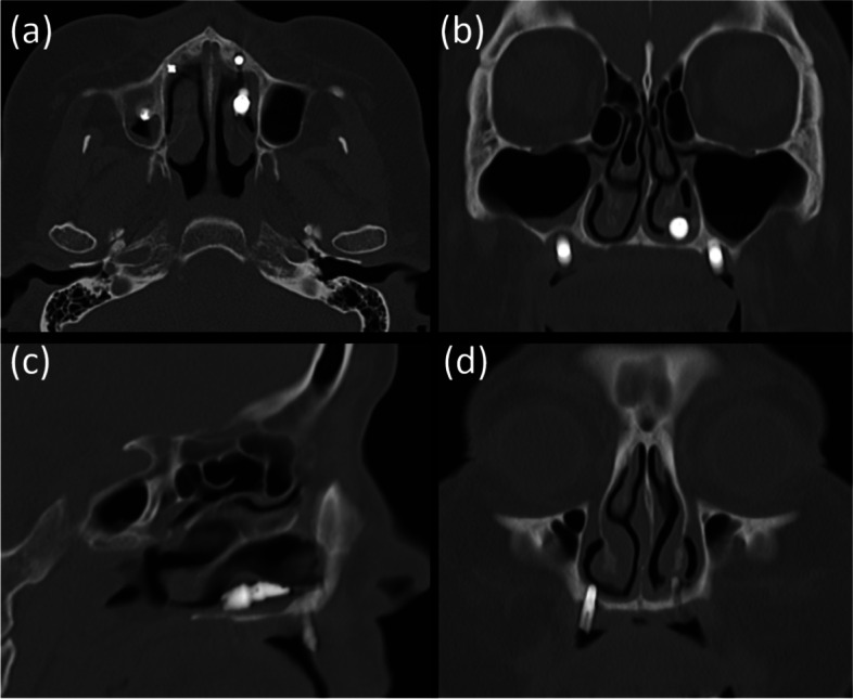
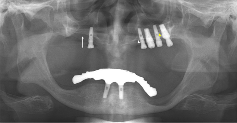
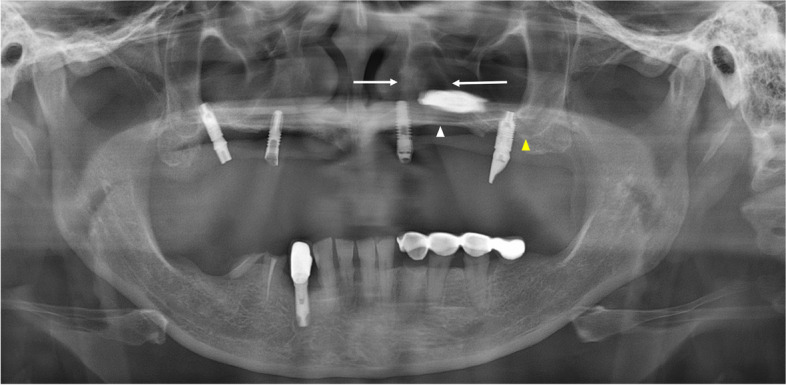
The nasal cavity is an important landmark when considering implant insertion into the anterior region of the maxillary arch. The perforation of implants into the nasal cavity may cause complications, such as implant migration, inflammation, or changes in nasal airflow; thus, precise assessment of the nasal cavity is mandatory.Three cases of nasal cavity perforation by dental implants are presented, including one case of implant fixture migration into the nasal cavity. On panoramic radiographs of the patients, the following common features were observed: the horizontal radiopaque line of the hard palate was observed to be inferior to or similar to that of the antral floor and the bone between the lateral wall of the nasal cavity and the medial wall of the maxillary sinus was emphasized in a triangular shape.When the maxillary sinus is small and alveolar bone resorption is severe, panoramic evaluation may cause overestimation of the available residual bone, particularly in the maxillary canine/premolar region. Therefore, the residual bone should be reevaluated three-dimensionally to measure the exact bony shape and volume.
 求助内容:
求助内容: 应助结果提醒方式:


