Ghazal Mansouri, Leila Allahqoli, Hamid Salehiniya, Ibrahim Alkatout
{"title":"40岁女性外生殖器蝇蛆病一例。","authors":"Ghazal Mansouri, Leila Allahqoli, Hamid Salehiniya, Ibrahim Alkatout","doi":"10.1155/2023/5579531","DOIUrl":null,"url":null,"abstract":"<p><p>Human myiasis is an infestation produced by fly larvae invading the tissues. We present a case of a 40-year-old virgin woman with vulvar myiasis. She reported at the gynecology clinic with a bloody discharge, severe pain, and swelling of the genital area for six days. Her menstrual history revealed the use of folded clothes. She had no specific gynecological disease. At the examination of the external genitalia, a tender mass measuring 6 cm × 4 cm and an ulcer measuring 1 cm × 1 cm on the surface of the labia majora were found. The patient was hospitalized. Serology, blood, and urine tests were requested; all laboratory tests were normal. The patient was transferred to the operating room (OR) with the diagnosis of necrotizing fasciitis. In the OR, we performed a longitudinal incision on the mass and removed nearly 30 visible maggots. After washing with normal saline, the patient was transferred to the ward without wound suturing. Debridement of the necrotic vulvar mass along with daily washing was performed for 7 days. The wound was sutured on the seventh day at the OR. Antibiotic therapy was continued for 4 days, and the patient was discharged with normal laboratory tests on the eleventh day after admission. We believe that poor sanitary hygiene was the cause of vulvar myiasis in our patient. We conclude that appropriate measures must be taken to reduce the risk of human myiasis, especially in tropical rural regions.</p>","PeriodicalId":9610,"journal":{"name":"Case Reports in Obstetrics and Gynecology","volume":"2023 ","pages":"5579531"},"PeriodicalIF":0.8000,"publicationDate":"2023-01-01","publicationTypes":"Journal Article","fieldsOfStudy":null,"isOpenAccess":false,"openAccessPdf":"https://www.ncbi.nlm.nih.gov/pmc/articles/PMC10432004/pdf/","citationCount":"0","resultStr":"{\"title\":\"External Genitalia Myiasis in a 40-Year-Old Woman.\",\"authors\":\"Ghazal Mansouri, Leila Allahqoli, Hamid Salehiniya, Ibrahim Alkatout\",\"doi\":\"10.1155/2023/5579531\",\"DOIUrl\":null,\"url\":null,\"abstract\":\"<p><p>Human myiasis is an infestation produced by fly larvae invading the tissues. We present a case of a 40-year-old virgin woman with vulvar myiasis. She reported at the gynecology clinic with a bloody discharge, severe pain, and swelling of the genital area for six days. Her menstrual history revealed the use of folded clothes. She had no specific gynecological disease. At the examination of the external genitalia, a tender mass measuring 6 cm × 4 cm and an ulcer measuring 1 cm × 1 cm on the surface of the labia majora were found. The patient was hospitalized. Serology, blood, and urine tests were requested; all laboratory tests were normal. The patient was transferred to the operating room (OR) with the diagnosis of necrotizing fasciitis. In the OR, we performed a longitudinal incision on the mass and removed nearly 30 visible maggots. After washing with normal saline, the patient was transferred to the ward without wound suturing. Debridement of the necrotic vulvar mass along with daily washing was performed for 7 days. The wound was sutured on the seventh day at the OR. Antibiotic therapy was continued for 4 days, and the patient was discharged with normal laboratory tests on the eleventh day after admission. We believe that poor sanitary hygiene was the cause of vulvar myiasis in our patient. We conclude that appropriate measures must be taken to reduce the risk of human myiasis, especially in tropical rural regions.</p>\",\"PeriodicalId\":9610,\"journal\":{\"name\":\"Case Reports in Obstetrics and Gynecology\",\"volume\":\"2023 \",\"pages\":\"5579531\"},\"PeriodicalIF\":0.8000,\"publicationDate\":\"2023-01-01\",\"publicationTypes\":\"Journal Article\",\"fieldsOfStudy\":null,\"isOpenAccess\":false,\"openAccessPdf\":\"https://www.ncbi.nlm.nih.gov/pmc/articles/PMC10432004/pdf/\",\"citationCount\":\"0\",\"resultStr\":null,\"platform\":\"Semanticscholar\",\"paperid\":null,\"PeriodicalName\":\"Case Reports in Obstetrics and Gynecology\",\"FirstCategoryId\":\"1085\",\"ListUrlMain\":\"https://doi.org/10.1155/2023/5579531\",\"RegionNum\":0,\"RegionCategory\":null,\"ArticlePicture\":[],\"TitleCN\":null,\"AbstractTextCN\":null,\"PMCID\":null,\"EPubDate\":\"\",\"PubModel\":\"\",\"JCR\":\"Q4\",\"JCRName\":\"OBSTETRICS & GYNECOLOGY\",\"Score\":null,\"Total\":0}","platform":"Semanticscholar","paperid":null,"PeriodicalName":"Case Reports in Obstetrics and Gynecology","FirstCategoryId":"1085","ListUrlMain":"https://doi.org/10.1155/2023/5579531","RegionNum":0,"RegionCategory":null,"ArticlePicture":[],"TitleCN":null,"AbstractTextCN":null,"PMCID":null,"EPubDate":"","PubModel":"","JCR":"Q4","JCRName":"OBSTETRICS & GYNECOLOGY","Score":null,"Total":0}
引用次数: 0
摘要
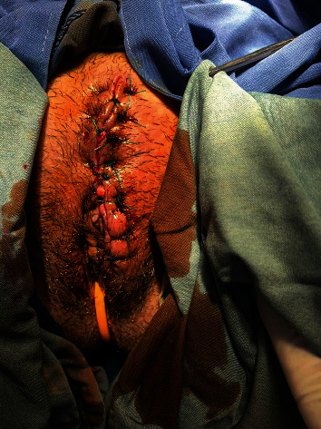
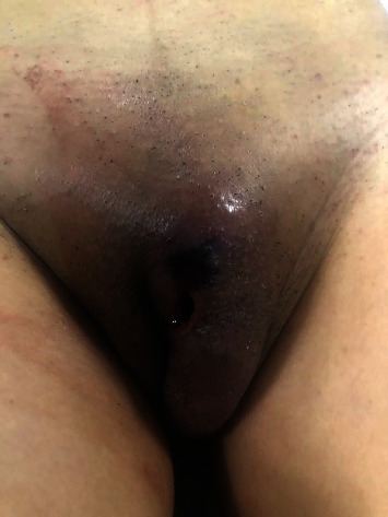
蝇蛆病是一种由蝇幼虫侵入人体组织而引起的疾病。我们提出一个40岁的处女女性外阴丝虫病的情况。她在妇科诊所报告,分泌物带血,剧烈疼痛,生殖器区域肿胀6天。她的经期记录显示她经常叠衣服。她没有特殊的妇科疾病。检查外生殖器时,发现大阴唇表面有6 cm × 4 cm的压痛肿块和1 cm × 1 cm的溃疡。病人被送进医院。要求进行血清学、血液和尿液检查;所有实验室检查都正常诊断为坏死性筋膜炎,转入手术室。在手术室里,我们在肿块上做了一个纵向切口,切除了近30个可见的蛆。用生理盐水冲洗后,未缝合伤口转移至病房。对坏死外阴肿块进行清创,并每日清洗7天。第7天在手术室缝合伤口。抗生素治疗持续4 d,患者入院后第11天实验室检查正常出院。我们认为恶劣的卫生条件是导致我们患者外阴丝虫病的原因。我们的结论是,必须采取适当措施来减少人蝇病的风险,特别是在热带农村地区。
External Genitalia Myiasis in a 40-Year-Old Woman.
Human myiasis is an infestation produced by fly larvae invading the tissues. We present a case of a 40-year-old virgin woman with vulvar myiasis. She reported at the gynecology clinic with a bloody discharge, severe pain, and swelling of the genital area for six days. Her menstrual history revealed the use of folded clothes. She had no specific gynecological disease. At the examination of the external genitalia, a tender mass measuring 6 cm × 4 cm and an ulcer measuring 1 cm × 1 cm on the surface of the labia majora were found. The patient was hospitalized. Serology, blood, and urine tests were requested; all laboratory tests were normal. The patient was transferred to the operating room (OR) with the diagnosis of necrotizing fasciitis. In the OR, we performed a longitudinal incision on the mass and removed nearly 30 visible maggots. After washing with normal saline, the patient was transferred to the ward without wound suturing. Debridement of the necrotic vulvar mass along with daily washing was performed for 7 days. The wound was sutured on the seventh day at the OR. Antibiotic therapy was continued for 4 days, and the patient was discharged with normal laboratory tests on the eleventh day after admission. We believe that poor sanitary hygiene was the cause of vulvar myiasis in our patient. We conclude that appropriate measures must be taken to reduce the risk of human myiasis, especially in tropical rural regions.

 求助内容:
求助内容: 应助结果提醒方式:


